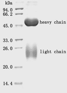
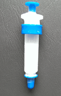
LifeTein Protein G Agarose consists of recombinant Protein G that has been covalently immobilized at high density onto high-quality crosslinked 6% beaded agarose. Protein G is especially suited for use with mouse antibodies in addition to most IgG isotypes from human, goat, cow and sheep sera, including human IgG3 and mouse IgG1 isotypes.
Protein G is a bacterial cell wall protein original from group G Streptococcus and now produced as a recombinant in E. coli. Recombinant Protein G has a mass of approximately 31 to 34 kDa by SDS-PAGE. IgG-binding function is optimal at pH 5 but also occurs efficiently in near-neutral conditions (pH 7.0 to 7.2). Protein G binds to most mammalian immunoglobulins primarily through their Fc regions.
Compared to Protein A, Protein G binds a broader spectrum of IgG subclasses from human, mouse and rat serum. Protein G exhibits stronger binding to human IgG3, mouse IgG1 and all three isotypes of Rat IgG. However Protein G binds more weakly than Protein A to pig, guinea pig, dog and cat IgG.
Protocol:
Protein G Binding buffer: 20 mM sodium phosphate, 150 mM NaCl, pH 7.4
Protein G Elution buffer: 100 mM Glycine, pH 3.0
Protein G Neutralization buffer: 1M Tris buffer, pH 7.5-9
- Wash the prepacked column with 5~10 column volumes of distilled water to remove 20% ethanol.
- Equilibrate the column with 5~10 column volumes of binding buffer.
- 1:10 dilution of serum with binding buffer. Filtrate the diluted serum through a 0.45 μm filter and load the sample.
- Wash with 10 column volumes of binding buffer.
- Elute with 5 column volumes of elution buffer and neutralize collect fractions with neutralization buffer.
- After each separation cycle, regenerate the resin by washing with approximately 3~5 column volumes of
0.1 M citrate buffer (pH 3.0).
- Confirm the purity of the collected antibody by SDS-PAGE analysis.
- Purification capacity: ≥30 mg of rabbit IgG per ml of recombinant protein G agarose gel.
 
|  https://www.lifetein.com
100 Randolph Road, Suite 2D,
Somerset
USA
New Jersey
08873
 https://www.lifetein.com
100 Randolph Road, Suite 2D,
Somerset
USA
New Jersey
08873