Unusual Amino Acids: Naphthylalanine

1-naphthylalanine
1-naphthylalanine (1-Nal)

Unusual amino acids represent a fascinating frontier in biochemistry and molecular engineering, offering functionalities beyond the 20 canonical proteinogenic amino acids. Among these, naphthylalanine (Nal) stands out for its unique structural and photophysical properties. Characterized by a naphthalene ring system, this non-natural amino acid exists in two primary isomeric forms: 1-naphthylalanine (1-Nal) and 2-naphthylalanine (2-Nal), distinguished by the attachment position of the naphthyl group to the alanine backbone. Its bulky aromatic side chain enhances hydrophobicity and steric influence, making it invaluable for probing protein folding, receptor-ligand interactions, and fluorescence-based applications. Consequently, Nal has become a cornerstone in peptide engineering and synthetic biology.


Key Takeaways

  • Structural versatility: Nal’s isomers (1-Nal and 2-Nal) provide distinct steric and electronic profiles for peptide design.
  • Bioconjugation compatibility: Amenable to solid-phase peptide synthesis (SPPS) using Fmoc- or Boc-protected derivatives.
  • Research utility: Critical for studying protein interactions, enzyme specificity, and cellular uptake mechanisms.

Structural and Chemical Properties

Molecular Architecture

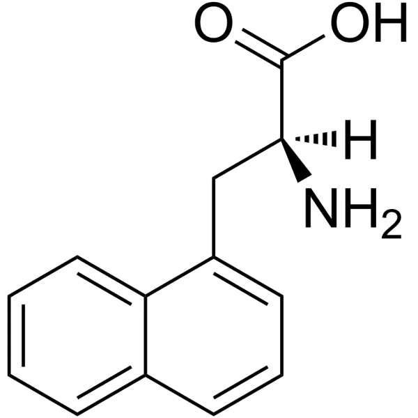
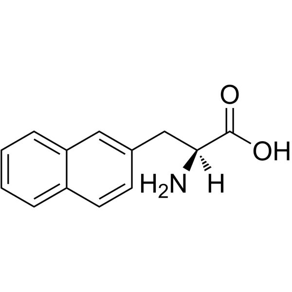
Naphthylalanine (C₁₃H₁₃NO₂) features a naphthalene moiety fused to the β-carbon of alanine. The 1-Nal isomer (CAS 55516-54-6) attaches the naphthyl group at the 1-position, while 2-Nal (CAS 58438-03-2) attaches it at the 2-position. This difference significantly impacts their chemical behavior: 1-Nal exhibits greater steric hindrance and a higher melting point compared to 2-Nal. Both isomers are soluble in organic solvents (e.g., DMSO, chloroform) but exhibit limited water solubility, necessitating tailored buffer conditions for biological assays.

Spectral and Electronic Traits

The extended π-conjugation of the naphthalene ring confers intrinsic fluorescence, with absorption/emission profiles suitable for UV-Vis detection. Furthermore, its hydrophobicity enhances membrane permeability, making it ideal for cell-penetrating peptide designs.

Find out more about peptide synthesis here.


Applications in Biochemical Research

Peptide Therapeutics and Drug Design

Nal’s hydrophobicity and stability enhance peptide-drug pharmacokinetics. It is incorporated into peptidomimetics targeting enzymes or receptors. Notably, Nal derivatives bind the Salmonella typhimurium OppA transporter, revealing pathways for antimicrobial development.

2-naphthylalanine
2-naphthylalanine (2-Nal)

Synthesis and Conjugation Methods

Solid-Phase Peptide Synthesis (SPPS)

Nal is incorporated into peptides using Fmoc- or Boc-protected precursors (e.g., Fmoc-1-Nal-OH, CAS 96402-49-2). LifeTein’s SPPS protocols achieve high-purity (>95%) Nal-labeled peptides, even for highly hydrophobic sequences up to 68 amino acids. Critical considerations include:

  • Spacer integration: Aminohexanoic acid (Ahx) spacers prevent steric hindrance during dye conjugation.
  • Orthogonal protection: Boc groups preserve side-chain functionality during fluorescent labeling.

Find out about high-speed RUSH synthesis.


Future Directions

Ongoing innovations include genetic code expansion to incorporate Nal in vivo via stop codon suppression. Additionally, multiphoton FRET using Nal’s UV-shifted spectra could enable deeper tissue imaging.


Frequently Asked Questions (FAQ)

What distinguishes 1-Nal from 2-Nal?

The attachment position of the naphthyl group: 1-Nal links at the naphthalene’s 1-position, causing greater steric hindrance, while 2-Nal links at the 2-position, offering milder steric effects 48.

Why use Nal instead of phenylalanine in peptide design?

Nal’s larger aromatic surface enhances hydrophobic interactions and fluorescence quenching efficiency, improving sensitivity in FRET and protein-binding studies 39.

Is Nal suitable for cell-penetrating peptides (CPPs)?

Absolutely. Its hydrophobicity enhances membrane permeability, and LifeTein couples it to TAT or R8 CPPs for intracellular delivery studies.

Fluorescent Labeling Pairs with EDANS and DABCYL

EDANS and DABCYL
EDANS (1,5-EDANS)

Fluorescence Resonance Energy Transfer (FRET) technology has revolutionized molecular detection by enabling real-time monitoring of biomolecular interactions and enzymatic activity. Among the most effective FRET pairs, the EDANS and DABCYL combination stands out for its exceptional quenching efficiency and widespread application in protease research. This fluorophore-quencher system operates through non-radiative energy transfer, where the excited-state energy of EDANS (donor) is absorbed by DABCYL (acceptor) when positioned within 10–100 Å. Consequently, the intact molecular construct exhibits minimal fluorescence, while proteolytic cleavage or conformational separation generates a robust, quantifiable signal. This mechanism provides unparalleled sensitivity for tracking dynamic biochemical processes in complex biological environments.


Key Takeaways

  • EDANS/DABCYL exhibits optimal spectral overlap with excitation at 341 nm and emission at 471 nm for EDANS, and DABCYL absorption at 453 nm.
  • Applications span protease activity profilingHIV protease inhibitor screening, and real-time enzymatic kinetics.
  • Solid-phase synthesis via Fmoc-Glu(EDANS)-OH and Fmoc-Lys(DABCYL)-OH enables precise incorporation into peptide sequences.

Spectral Properties and Quenching Mechanism

Molecular Characteristics

EDANS (5-((2-Aminoethyl)amino)naphthalene-1-sulfonic acid) functions as the donor fluorophore with excitation/emission maxima at 341/471 nm. Its extended conjugated system provides high quantum yield, while its sulfonate group enhances water solubility. Conversely, DABCYL (4-((4-(Dimethylamino)phenyl)azo)benzoic acid) serves as a non-fluorescent “dark quencher” with broad absorption (λmax 453 nm). This spectral profile allows near-complete overlap between EDANS emission and DABCYL absorption, fulfilling the Förster radius requirement for efficient energy transfer (typically 3–5 nm). Furthermore, DABCYL’s lack of intrinsic fluorescence eliminates background noise, significantly enhancing signal-to-noise ratios in detection assays.

Intramolecular Quenching Dynamics

The quenching efficiency of this pair stems from dipole-dipole coupling, where electronic excitation energy transfers from EDANS to DABCYL without photon emission. Critically, this transfer rate decays inversely with the sixth power of the distance between fluorophores, making the system exquisitely sensitive to molecular separation. In protease substrates, the spacer sequence between EDANS and DABCYL adopts an extended conformation to maximize quenching. Upon enzymatic cleavage, the fragments diffuse apart, disrupting energy transfer and permitting EDANS fluorescence recovery. This mechanism enables continuous real-time monitoring of enzymatic kinetics without secondary detection reagents.

Find out more about fluorescent peptides here.


Applications in Biochemical Research

Protease Activity Profiling

The EDANS/DABCYL pair has become indispensable for protease specificity studies, particularly for HIV-1 protease research. Researchers design peptide substrates mimicking viral polyprotein cleavage sites, flanked by EDANS (N-terminus) and DABCYL (C-terminus). In intact substrates, fluorescence remains quenched >95%. However, protease cleavage yields fluorescence increases proportional to enzyme concentration, permitting detection limits in the nanomolar range. This approach facilitates rapid screening of protease inhibitors and kinetic characterization of mutant enzymes.

Real-Time Cellular Imaging

Although chemical dyes face challenges in live-cell imaging due to cytotoxicity, modified EDANS/DABCYL constructs enable intracellular protease mapping. When conjugated to cell-penetrating peptides (CPPs), these substrates can monitor caspase activity during apoptosis or viral infection cycles. Nevertheless, researchers must optimize delivery vehicles and exposure parameters to minimize phototoxicity, as the EDANS excitation wavelength (341 nm) approaches the UV range. Mitigation strategies include pulsed illumination, oxygen scavenging, and serum-free media to reduce background fluorescence.


Practical Implementation Considerations

EDANS and DABCYL
DABCYL acid

Solid-Phase Synthesis Strategies

Incorporating EDANS/DABCYL into peptides traditionally faced synthetic hurdles due to EDANS’s poor nucleophilicity and DABCYL’s steric constraints. Modern approaches utilize pre-derivatized building blocks:

  • Fmoc-Glu(EDANS)-OH: Incorporates EDANS via glutamic acid side chain
  • Fmoc-Lys(DABCYL)-OH: Anchors DABCYL to lysine residues

Comparison with Alternative FRET Pairs

Performance Metrics

FRET PairFörster Distance (Å)Fluorescence EnhancementPrimary Applications
EDANS/DABCYL33–4140-foldProtease substrates, nucleic acid probes
Mca/Dnp28–3225-foldMetalloprotease assays
Cy3/Cy5>5015-foldProtein interaction studies
FITC/TAMRA49–5530-foldCell imaging

While Cy3/Cy5 offers superior Förster distances (>50Å), its fluorescence enhancement remains lower due to acceptor emission. Conversely, Mca/Dnp provides higher temporal resolution in zinc-dependent proteases but suffers from lower photostability. Consequently, EDANS/DABCYL remains the gold standard for sensitive endoprotease detection where cleavage sites permit optimal fluorophore spacing.

Emerging Alternatives

ATTO 550 has emerged as a photostable alternative to Cy3 in FRET applications, exhibiting higher brightness and reduced cytotoxicity. LifeTein’s ATTO conjugation services now offer this dye as a replacement for traditional EDANS in multiplexed assays. Nevertheless, DABCYL derivatives maintain dominance as dark quenchers due to their broad absorption spectra and commercial availability.

Find out more about peptide synthesis here.


Frequently Asked Questions

Why is EDANS/DABCYL preferred over other FRET pairs for protease assays?

The combination provides exceptional quenching efficiency (>95%) and high signal-to-noise ratios upon cleavage due to DABCYL’s non-fluorescent nature. Its spectral overlap permits 40-fold fluorescence enhancement, significantly outperforming Mca/Dnp (25-fold) and FITC/TAMRA (30-fold) pairs in sensitivity.

Can DABCYL quench fluorophores beyond EDANS?

Yes. DABCYL’s broad absorption spectrum (400–500 nm) enables efficient quenching of FAMTET, and Mca. However, its extinction coefficient is highest near 453 nm, making EDANS (emission 471 nm) the optimal partner.

What are key applications beyond protease detection?

This FRET pair enables:

  • Nucleic acid hybridization probes (molecular beacons)
  • Protein conformational change sensors
  • Antibody-epitope binding kinetics
  • High-throughput drug screening platforms

How does this pair compare to Cy3/Cy5 in live-cell imaging?

While Cy3/Cy5 offers superior photostability for longitudinal studies, EDANS/DABCYL provides higher sensitivity for endpoint assays. However, EDANS’s UV excitation (341 nm) increases phototoxicity risks, making Cy3/Cy5 preferable for extended live-cell observation.