About LifeTein Peptide

The custom peptide synthesis service and custom antibody product service company.

Synthetic Scorpion Toxin Peptides: A New Frontier in Chronic Pain Management by LifeTein

Synthetic Scorpion Toxin Peptides
Synthetic Wasabi Receptor Toxin

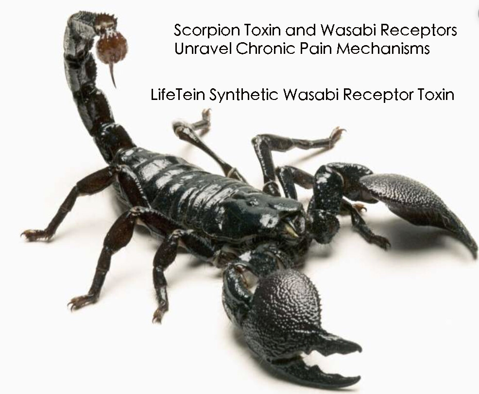
Researchers at the University of California, San Francisco (UCSF), in collaboration with LifeTein, have made a groundbreaking discovery in the field of pain management. LifeTein’s expertise in peptide synthesis was crucial in developing synthetic scorpion toxin peptides that specifically target the “wasabi receptor,” a key player in the body’s response to certain types of pain.

The wasabi receptor, scientifically known as TRPA1, is an ion channel protein that triggers the familiar sinus-clearing or eye-watering sensation experienced when consuming wasabi or cutting onions. This receptor is also implicated in the perception of chronic pain.

The focus of this research is a peptide derived from scorpion toxin, referred to as WaTx. Remarkably, WaTx, synthesized by LifeTein, can activate the TRPA1 receptor, mimicking the pain response to irritants. Unlike other molecules, WaTx has the unique ability to penetrate cell membranes directly, bypassing the need for channel proteins. This property makes it an invaluable tool for studying chronic pain and inflammation.

In addition to its research applications, WaTx holds promise for the development of new, non-opioid pain therapies. It has been observed to induce pain and pain hypersensitivity without causing neurogenic inflammation, a common side effect of many pain treatments.

Expanding the Horizon: Spider Venom and Chronic Pain

Further expanding on this concept, a study titled “Identification and Characterization of ProTx-III [μ-TRTX-Tp1a], a New Voltage-Gated Sodium Channel Inhibitor from Venom of the Tarantula Thrixopelma pruriens” delves into the potential of spider venoms in pain management. This study, conducted by F. C. Cardoso and colleagues, discovered a novel inhibitor, μ-TRTX-Tp1a (Tp1a), from the venom of the Peruvian green-velvet tarantula. Tp1a selectively inhibits human NaV1.

7 channels, which are key contributors to pain perception.

The study found that Tp1a, both in its recombinant and synthetic forms, preferentially targets NaV1.7 channels, offering a new avenue for analgesic drug development. Unlike many other spider toxins affecting NaV channels, Tp1a does not significantly alter the voltage dependence of activation or inactivation of these channels. This unique feature of Tp1a was demonstrated to be effective in reversing spontaneous pain in animal models.

The structural analysis of Tp1a revealed an inhibitor cystine knot motif, common in spider toxins but with distinct pharmacological properties that could be crucial in developing more selective and potent treatments for chronic pain.

Conclusion

The research at UCSF, along with the findings on spider venom peptides and the significant contributions of LifeTein in peptide synthesis, represents a significant step forward in understanding and potentially treating chronic pain. These discoveries highlight the vast potential of natural toxins in medical research, offering hope for more effective and safer pain management strategies in the future.

Reference:

  • Lin King, J. V., Emrick, J. J., Kelly, M. J. S., Herzig, V., King, G. F., Medzihradszky, K. F., & Julius, D. (2019). A Cell-Penetrating Scorpion Toxin Enables Mode-Specific Modulation of TRPA1 and Pain. Cell. doi:10.1016/j.cell.2019.07.014

LifeTein peptide FLAG(GS)HA: DYKDDDDK-GGGGS-YPYDVPDYA-NH2 helped discover insulin-like peptide6, Dilp6, in regulating growth in fruit flies Drosophila

FLAG and HA tagged IGF1

In humans, liver-derived insulin-like growth factor (IGF1) drives postnatal growth. Early childhood infection of E. coli, Campylobacter spp., even asymptomatic, reduces IGF1 level and restricts early-childhood growth. Does the pathogen-induced Toll-like innate immune signaling contribute to growth restriction? To answer the question, the researchers examined a corresponding pathway in fruit flies.

LifeTein’s Peptide: FLAG(GS)HA

In fruit flies, Dilps (Drosophila insulin-like peptides) drive their growth, for example, the growth rate of imaginal discs which give rise to adult structures such as wings. Dilps share homology with insulin and IGF1, and they bind to the insulin receptor. Dilp6 is produced by fat body, an organ for nutrient storage and immune functions.

The researchers found Dilp6 is a selective target of Toll signaling in the fat body, an innate immune response from bacterial infections. They also found that Toll signaling reduces Dilp6 transcripts, and dramatically suppresses circulatory Dilp6 levels, and restricts whole-body growth. Restoring Dilp6, on the other hand, rescues growth and viability in fruit flies even with active Toll signaling.

LifeTein’s peptide FLAG(GS)HA was used as a standard in ELISA to quantify Dilp6 in fruit fly hemolymph samples. Here, Dilp6 was tagged with FLAG and HA because of FLAG- and HA-tagged Dilp6HF allele from CRISPR/CAS9. In this ELISA assay, the plate wells were coated with anti-FLAG antibody, then FLAG(GS)HA or fruit fly hemolymph sample were added to the wells. FLAG(GS)HA and FLAG- and HA-tagged Dilp6 were quantified by anti-HA-Peroxidase 3F10 antibody and subsequent chromogenic reaction. For more details of the method, see the section “Hemolymph Dilp6 measurements by ELISA” in the link.

https://www.sciencedirect.com/science/article/pii/S2211124719309052

Peptide Synthesis Home Page

Our Services:

COVID-19 Services & Products

Custom Antibody Services

Rush Peptide Synthesis

Peptide Nucleic Acids (PNAs)

Custom Peptide Synthesis Services

Gene Synthesis Service

Custom Chemical Synthesis

Other Posts:

Smaller Ions Stabilize β-sheets

Synthetic Scorpion Toxin Peptides for Chronic Pain

Self-Assembling Peptide Hydrogels As a Drug Delivery System

LifeTein’s Braftide & Cancer Therapy

Ionic Magic: Small Ions Transform Peptides into Stable β-Sheets

Peptide amphiphiles are composed of hydrophobic alkyl tails and peptide regions designed to self-assemble into cylindrical supramolecular nanofibers in solution. While hydrogen bonds form some β-sheets between short β-strands (2 or 3 residues), others are formed by extended-strands.

Smaller Ions Stabilize β-sheets

The strongly-hydrated ions (F- and Cl-) are more attracted to the positively charged lysine residues on the surface of the peptide nanofiber. When peptide residues form β-sheets, an F- or Cl- ion forms a salt bridge between the side chains of lysine residues from two neighboring peptide amphiphile chains. The salt bridge stabilizes the peptide by bringing the backbones closer, resulting in a transition from random coil to extended β-sheets structures. The smaller ions (F- and Cl-, 50mM NaF and NaCl solutions) tend to stabilize β-sheets slightly better compared to the larger ions (I-, Br-).

So, the self-assembly of peptide amphiphiles into supramolecular nanofibers can be regulated by modifying the salt solution.

Reference: doi.org/10.1021/acs.jpcb.9b05532

Self-Assembling Peptide Hydrogel as a Versatile Drug Delivery Platform

Peptide Hydrogel

Hydrogels with a capacity to absorb and hold water within a porous, swelled structure make it a great candidate as a drug delivery system due to their broad range of physical properties as well as chemical adaptability. For example, a positively charged polypeptide (poly-l-lysine, PLL) coupling to a self-assembling dipeptide (Fmoc-FF) leads to the formation of hydrogels with rheological properties suitable for injection.

Self-Assembling Peptide Hydrogels As a Drug Delivery System

Hydrogenation via self-assembly is a hierarchical process. The hydrogels can be easily prepared via simple mixing and incorporated with various stoichiometric ratios of peptides without any additional synthetic processes. Peptides can form hydrogels by multiple non-covalent interactions. It was found that PTZ-Gly-Phe-Phe-Tyr can form gels at ultra-low concentrations of 0.01 wt.%. The π-π stacking may be another driving forces in the self-assembly to form the final hydrogel structure. The N-cadherin mimic peptide (CLRAHAVDIN) and TGF-β1 mimic peptide (CESPLKRQ) synthesized by LifeTein were used to form an injectable 3D hydrogel. Increased retention of stem cells by using an injectable hydrogel has resulted in successful tissue engineering outcomes.

Another efficient method to facilitate hydrogel formation is based on the electrostatic attraction of oppositely charged peptides. The order of amino acids is of great importance for hydrogenation. Self-assembling beta-hairpin peptides, with high arginine content, exhibited extremely good performance in killing bacteria. The peptide hydrogels also have been demonstrated to be used as wound healing agents and other therapeutic instruments under different conditions. Curcumin could be encapsulated into hairpin hydrogels as an injectable agent for localized delivery.

Peptide Synthesis Home Page

Our Services:

COVID-19 Services & Products

Custom Antibody Services

Rush Peptide Synthesis

Peptide Nucleic Acids (PNAs)

Custom Peptide Synthesis Services

Gene Synthesis Service

Custom Chemical Synthesis

Other Posts:

Smaller Ions Stabilize β-sheets

Synthetic Scorpion Toxin Peptides for Chronic Pain

LifeTein’s Braftide & Cancer Therapy

How to Identify a PD-L1 Binding Peptide for Determination of PDL1 Expression in Tumors

PD-L1 Binding Peptides

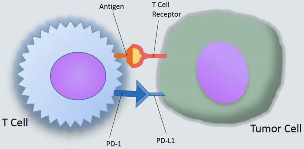
Blocking the interaction between Programmed Death Ligand 1 (PD-L1) and its receptor, PD-1, is an effective method of treating many types of cancers.

  1. Identifying the PD-L1 binding peptide and mock peptide: Select from a library of peptides for stability and high affinity.
  2. Fluorescent target peptide-Cy5 as Marker for Flow Cytometry.
  3. Detection of PD-L1 in circulating tumor cells using the Cy5 or Cy7 (infrared) labeled peptides.
  4. Biotinylated peptides to detect PD-L1 in sample tissues. The streptavidin-HRP are used.
  5. Fluorescent Cy5-labeled peptides detects PD-L1 in sample tissues.

By utilizing a peptide-based approach, it is possible to detect all levels of PD-L1 with high sensitivity and specificity.

Reference:

https://www.nature.com/articles/s41598-017-10946-2

How to screen small peptide ligands for targeting receptors in cancer cells?

Neoepitopes as cancer immunotherapy targets: key challenges and opportunities
Neoepitopes as cancer immunotherapy targets
  1. Design a biotin-labeled peptide library: overlapping or Alanine scan
  2. The peptide library is coated on the avidin-coated 96 well plates.
  3. The Peptide library is used to evaluate the binding of each peptide to different cell lines using CyQUANT Cell Proliferation Assays.
  4. Flow cytometry: Determine the cellular uptake of selected FITC-labeled peptides.
  5. Competitive assay: The cells are incubated with FITC-peptide with or without excess unlabeled peptide (100-fold)
  6. Finetune the peptide sequence: structure analysis, cyclization, and D amino acid modification.
  7. Find the promising receptor-specific cancer cell binding peptide that can be conjugated directly to a chemotherapeutic drug or to nanoparticles for targeted drug delivery to enhance the efficacy of chemotherapy for cancer treatment.

Reference:

https://www.nature.com/articles/s41598-019-38574-y

Synthetic Peptides as Protein Mimics in Biological Research

Synthetic Peptides as Protein Mimics

Synthetic peptides have proven an excellent type of molecule for the mimicry of protein sites. The modified peptides increase the proteolytic stability of the molecules, enhancing their utility for biological applications.

Toolbox for Peptide Synthesis: Non-Proteinogenic Amino Acids and Site-Selective Ligation

The long peptides can be synthesized by the ligation method. Amino acid derivatives with modified backbone length and side-chain orientation, such as d-amino acids, N-alkyl glycine monomers, or proteolytically stable amino acid derivatives can be introduced to the peptides.

Protein Secondary Structure Mimics: α-Helix Mimics, β-Sheet Mimics
Peptide chains can be organized into secondary structures, such as α-helices and β-sheets. Peptides that mimic α-helices and β-sheets of proteins are attractive targets for drug development and tools to explore protein binding mechanism.

The α-helical conformation of a peptide can be induced by adding covalent links between amino acid side chains at selected positions. These links can be formed by lactam and disulfide bridges, triazole-based linkages, and hydrocarbon staples.

In β-sheets, β-strands are connected via loops or turns. Methods to mimic turn structures include macrocyclization, dipeptide of d-proline and l-proline, or α-aminoisobutyric acid in combination with either a d-α-amino acid or an achiral α-amino acid. An example of stimuli-responsive peptides is the temperature-dependent formation of hydrogels by β-sheet peptides. The β-hairpin mimic undergoes gelation upon heating at 60°C, and is completely reversible while cooling.

Protein Mimics in Biomedical Research

Peptides mimicking the CHR region of gp41 were developed to inhibit the formation of the six-helical bundle. Peptides that mimic these receptors are useful tools to explore the details of virus infection mechanism, as well as to develop new drugs against HIV-1. Peptides that mimic the extracellular domains of seven transmembrane G protein-coupled receptors (GPCRs), which is composed of the N-terminus (NT) and the three extracellular loops (ECLs) were explored. Peptide Ac-RERF-NH2 has a high propensity to adopt an α-turn structure and could be a promising drug candidate against cancer.

The design of peptides as protein mimics has evolved as a promising strategy for the exploration of protein-protein interactions, as they are biocompatible, biodegradable, and functionally selective.

Explainer: Why peptides are the ‘next big thing’ in medical research?

Cell Penetrating Peptides for Drug Discovery

Biochemists are excited by the possibilities presented by peptides and proteins as pharmaceuticals because they so often mimic exactly the behavior of a natural ligand – the substance that interacts with the receptor on an enzyme or cell to cause a biological process.

Peptides are used for:
1. Self-Assembling Peptide Epitopes as Novel Platform for Anticancer Vaccination: OVA 250−264 and HPV16 E7 43−57.

2.Incorporation of DSPE-PEG and cRGD-modified DSPE-PEG molecules improves the biocompatibility and cellular uptake of the nanoprodrug platform: Click chemistry conjugation of peptides to PEGs

3. Designing Tracers for Imaging from Cyclic Peptides: ATTO, FITC, FAM, Cy3, Cy5, infrared C7

4. Co-delivery of tumor antigen and dual toll-like receptor ligands into dendritic cell: Cell Penetrating Peptides, HIV-TAT proteins, R8.

How to Design Peptide for Antimicrobial Applications

Antimicrobial Peptides

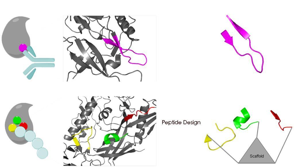
There are many ways to design improved peptides: site-directed mutagenesis, computational design approaches, synthetic libraries, template-assisted methodologies, and mechanism-based strategies.

  1. Site-directed mutagenesis:
    Adding, deleting, or replacing one or more amino acid residues are useful approaches to re-engineer a peptide. The alanine or lysine scanning are examples of valuable strategies, as they allow for coverage of all positions within a peptide chain, thus making it possible to analyze the effect of all amino acid side chains on structure and function.
  2. De novo design:
    Peptides present both basic and hydrophobic residues in a given sequence. The antimicrobial peptides favor an amphipathic structure. The helix-stabilizing residues such as leucine, alanine, valine, isoleucine, lysine, and arginine, or prototypical destabilizing residues such as proline and glycine are frequently introduced to manipulate the peptide structure. The design can also base on the native bioactive peptides as the template-assisted approach.
  3. Synthetic libraries:
    A synthetic combinatorial library is practical to scan for the active binding site. The positional scanning or iterative approaches are costly and time-consuming.
  4. Computational methods:
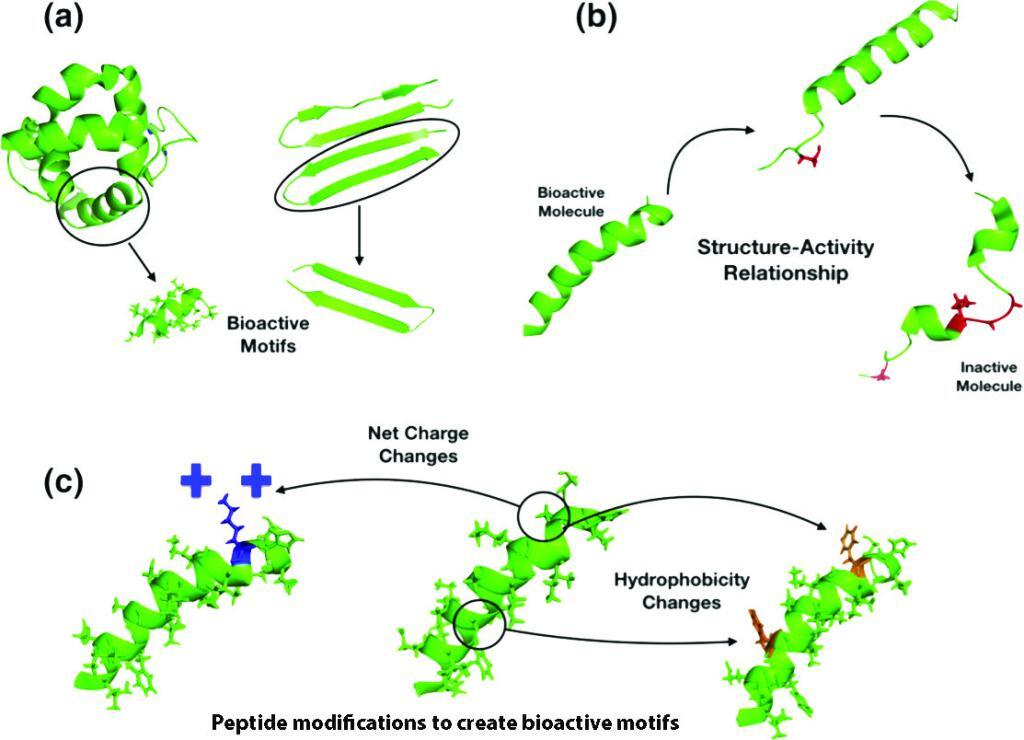
    The antimicrobial databases are widely available. The design can be based on the bioinformatics tools, statistical modeling, SAR studies, genetic algorithms or deep learning. The sequence motifs, structure, net charge, hydrophobicity, amphipathicity, and unnatural modifications are essential guidelines for the peptide design.

Perfluoroarene-based Peptide Macrocycles

Perfluoroarene-based Peptide Macrocycles

There are many methods to modify cysteine, which include alkylation, conjugate addition, oxidation, and reduction.

Recently, it was found that it is possible to arylate cysteine in the context of bioconjugation using sufficiently activated reagents. The cysteine perfluoroarylation can be used for mild functionalization of cysteine thiolate moieties.

This is a new and mild synthetic platform for cysteine arylation. The developed method operates at room temperature in polar organic solvents and shows excellent selectivity and functional group tolerance. As a result, one can utilize this approach towards stapling unprotected peptides, thereby positively altering their biological properties. It was demonstrated incorporation of a perfluoroaryl staple within the small tri-helical affibody protein.

Perfluoroarene-based peptide macrocycle is a stapling modification performed on a peptide sequence showed enhancement in binding, cell permeability, and proteolytic stability properties, as compared to the unstapled analog.

It was found that the utility of peptide macrocyclization through perfluoroaryl-cysteine chemistry to improve the ability of peptides to cross the blood−brain barrier. Multiple macrocyclic analogues of the peptide transportan-10 were investigated that displayed increased uptake in two different cell lines and improved proteolytic stability. The abiotic peptide macrocycles can exhibit significantly enhanced penetration of the brain.