Protein-protein interactions (PPI) are highly specific electrostatic attractions between protein structures. The interactions regulate cell function and influence physiology and development. Mass spectrometry is often used to detect protein-protein interactions. However, all proteomic screens are differential and require two samples such as IP and mock IP, POI and wildtype, or POI and knockout.
The peptide/ biotin-peptide or peptide/fluorescence-labeled peptide are excellent tools for studying protein-protein interactions. Check this link for details: Protein-Protein Interactions: Methods for Detection and Analysis. https://www.lifetein.com/Peptide_Modifications_biotinylation.html
The compounds that can modulate PPI are hard to discover because the proteins have multiple binding sites and the screening assays are not reliable. In many cases, two or more proteins may interact with one another and form a complex. The optical fluorescence-based methods such as the Cy5, Cy7, FAM, FITC, TAMRA-labeled peptides, or FRET assay are particularly useful in these circumstances. Click for more details: https://www.lifetein.com/Peptide-Synthesis-FITC-modification.html.
The interactions between a fluorescently labeled or intrinsically fluorescent sample and a binding patterner are measured during the application. The changes in intrinsic fluorescence from tryptophan and tyrosine residues in the protein can be measured, which indicates transitions in the protein’s folding state.
The scientists have been working on fusion-based bifunctional proteins in cancer immunotherapy. The bifunctional protein sent an apoptotic signal to the tumor cells and enhanced their killing. The click chemistry is the perfect tool for the drug-protein or protein-protein conjugation. The more we understand the natural receptor-ligand complex and how it might signal, the better we can guide the design of therapeutic agonists. Click here for the peptide conjugation details: https://www.lifetein.com/price_modification_labeling.html
Epitope mapping identifies antigen regions that serve as binding sites for antibodies. The overlapping linear peptides derived from the primary sequence of the antigen are frequently used for epitope screening. Individual peptides can be divided into several fragments that overlap. The resulting overlapping peptide libraries can then be used for processes including continuous and linear epitope mapping.
For example, to map the epitope of an antibody, a few overlapping fragments spanning the target regions are constructed in an expression vector. These constructs are transiently transfected in cells and whole cell lysates collected after 48 hours are subjected to Western blotting with the antibody. To further map the region of this fragment, a series of overlapping peptides are synthesized. These peptides and lysates from the cells expressing the target full-length gene or empty vector (negative) are performed by the dot blot.
Mapping epitopes quickly and accurately are challenging because the epitopes tend to be nonlinear on antigens. Combining binding specifically toward two distinct epitopes into a single molecule can significantly enhance the immunotherapeutic properties of monoclonal antibodies. Multivalent interactions are the most efficient at driving IgE receptor signaling pathways.
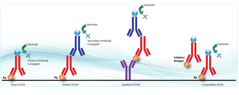
There are several useful tools for studying antigen-antibody interactions.
Use the MAPs as the immunogens. Multiple Antigenic Peptides (MAPs) are peptides that are branched artificially, in which Lys residues are used as the scaffolding core to support the formation of 8 branches with varying or the same peptide sequences. For example, one goal is to include different epitopes from different virus proteins in a single unit. The epitopes showing the right prediction of antigenicity and conserved in most serotypes of a virus are selected and assembled as the MAP.
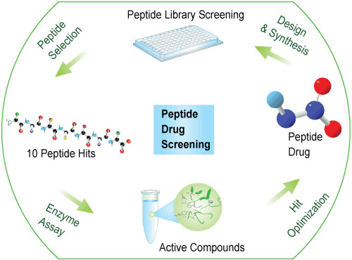
Screening combinatorial peptide libraries to optimize enzyme substrates and create high-affinity protein ligands. A critical biological application of custom peptide libraries is the characterization of the binding events that occur between specific proteins and their peptide ligands. A series of Overlapping Peptide, Truncation Peptide, Alanine Peptide Scanning, Scrambled Peptide, or Positional Peptide can be used for mapping and validating epitopes, the characterization of therapeutic antibodies, studying anti-antibody and neutralizing antibody actions in vitro.
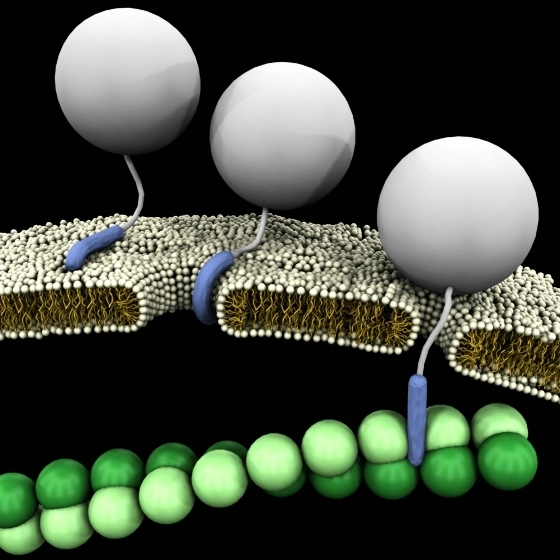
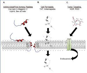
Tat, the transcription activator of the human immunodeficiency virus type 1 (HIV-1) viral genome, enters cells in a non-toxic and highly efficient manner. Tat is the first known cell-penetrating peptide.
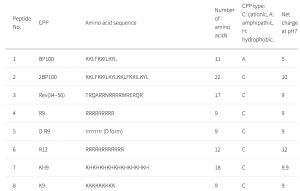
CPPs have been used as a carrier to deliver proteins or genes into cells and tissues. In this study, a CPP library composed of 55 CPPs was used to deliver genes into plant cells. Many CPPs showed efficient penetration into plant cells. The Lys-containing CPPs have higher penetration efficiency in the plant than in animal cells. This could be due to differences in lipid composition and surface charge of the cell membranes. No correlations were detected between the penetration efficiency and the cationic, amphipathic, or hydrophobic properties of peptides.
D-R9 is composed of D-form amino acids. D-R9 bound preferentially to the membrane and did not penetrate the cytosol or vacuole. In mammalian cells, poly-lysine-based CPPs are efficient and interact with membrane lipid head groups to induce wrapping of the membrane monolayers. Arg-rich peptides, such as the Tat peptide, are among the most efficient CPPs. Arg-rich CPPs may generate negative Gaussian membrane curvature to form pores or protrusions from endocytosis. The cell penetration efficiency of CPPs containing poly-Arg is higher than those containing poly-Lys. However, in a plant, Arg-rich CPPs are not the most efficient at penetrating plant cells.
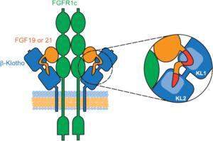
Endocrine fibroblast growth factors (FGFs) require Klotho transmembrane proteins as co-receptors to activate FGF receptor (FGFR) signaling.
A series of peptides were synthesized by LifeTein and used for the competition binding assay. Both the KL1 and KL2 domains of β-Klotho participate in ligand interaction. The FGF19 peptide was used for alanine scanning mutagenesis. It was found that a single amino acid mutation in either region was sufficient to abolish β-Klotho binding. FGF19 and FGF21 function through β-Klotho to regulate glucose and lipid metabolism.
How to perform the solid-phase binding assay
1. The 96-well plates were coated overnight at 4 °C with 2 µg/mL of antibody in PBS.
2. Plates were washed twice with PBST and blocked with 3% (w/v) BSA in PBS for 1.5 hours at room temperature.
3. The conditioned media containing β-Klotho were added to the plates and incubated for 1.5 hours at room temperature.
4. Plates were washed a few times.
5. The peptide mutation FGF21 and an anti-β-Klotho antibody were biotinylated with EZ-Link Sulfo-NHS-LC-Biotin at the indicated concentrations.
6. After washing, streptavidin-HRP was used for detection.
7. EC50 values were determined.
How to do a competition binding assay?
1. The WT and mutant peptides were custom synthesized and purified (>95% purity) by LifeTein.
2. Binding of FGF19 and FGF21 peptides to β-Klotho was assessed.
3. The β-Klotho ECD 6 × His, varying amounts of FGF19 and 21 peptides, and biotinylated human FGF19 or FGF21 protein were prepared.
4. The streptavidin donor beads and nickel chelate acceptor beads were added to the plates.
5. Plates were incubated for 3 hours at room temperature, protected from light, and read on the Plate Reader.
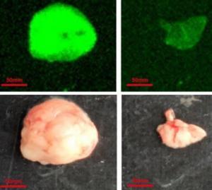
The folate receptor alpha (FRα) is highly expressed in ovarian cancer and not in normal tissues. An FRα binding peptide C7 (Met-His-Thr-Ala-Pro-Gly-Trp-Gly-Tyr-Arg-Leu-Ser, MHTAPGWGYRLS) was found to bind to FRα expressing cells. This tumor-targeting peptide was proved by both phage homing experiment and fluorescence imaging.
Tumor Targeting of Conjugated Synthetic Peptides
1. The FITC-conjugated peptide FITC-MHTAPGWGYRLS was dissolved in PBS.
2. The peptide was injected intravenously into a tumor-bearing nude mouse.
3. After 2 h, the tumor and other organ tissues were harvested and analyzed using a fluorescence imaging system.
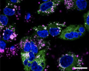
Cell internalization of Synthesized Peptide
1. Cells were seeded in 24 well plates containing coverslips and incubated for 24 hours in a medium with FBS.
2. FITC conjugated peptide was incubated with the cells for 4 hours at 37 °C.
3. The cells were washed once with PBS and fixed in 4% paraformaldehyde.
4. Cells were washed three times with PBS and stained with DAPI for 20 min at room temperature.
5. Internalized fluorescent signals were imaged with a confocal microscope.
This tumor-specific peptide could be a potent and selective ligand for FRα. It has a great potential for the delivery of cancer therapeutics or imaging agents to express tumors.
Cancer is expected to surpass the current number one, cardiovascular diseases by 2030 as the leading cause of death.
The targeting peptides can be considered as an alternative vehicle for the delivery of anti-cancer drugs because of their lower molecular weight and excellent tolerability by human bodies.
The following modification can prolong the half-life of the peptides from the degradation by blood proteases: forming cyclization within a peptide, blocking of the C- and N- terminus, replacement of standard L-type amino acids by their D-amino acid counterparts, using unnatural amino acids incompatible with endogenous proteases.
About targeting peptides: 1. Somatostatin (SST) derivatives: Binding of the natural ligand somatostatin peptide to the receptors leads to inhibition of overexpressed SSTR2 and 5 in breast cancer. For example, cyclic SSTR agonist octreotide (fCfDWKTCT), selectively binds SSTR2 and 5.
2. Peptide-derivatives of gastrin-releasing peptide (GRP)
The gastrin-releasing peptide receptor is associated with the prostate and breast cancer. It was found that bombesin (YQRLGNQWAVGHLM) and its derivatives could be used as targeting peptides for the detection of prostate cancer via PET and CT screening.
3. Peptides targeting tumor microenvironment
The glycoprotein prosaposin (PSAP) can inhibit metastases from breast and lung cancer in preclinical models. A cyclic PSAP peptide (DWLPK) could hinder metastatic spread and restrain tumor development. The synthetic antagonist of CXCR4 called NT21MP (LGASWHRPDKCCLGYQKRPLP) exhibits anti-tumor activities through decreased adhesion and migration of breast cancer cells. The peptide R (RACRFFC) targeting of CXCR4, showed capacities to remodel the tumor stroma.
4. Peptides targeting the tumor pH and temperature
The pHLIP (pH-Low Insertion Peptide), ACEQNPIYWARYADWLFTTPLLLLDLALLVDADET, can be inserted into cell membranes as an a-helix under low pH conditions. The pHLIP peptide increases the uptake of the peptide-coated drug to tumors compared to the naked particles. Thus, the local tumor microenvironment can be used to trigger peptide drug formulations to respond accordingly.
The elastin-like polypeptides (ELP) are conjugated to the cell-penetrating peptide Bac (RRIRPRPPRLPRPRPRPLPFPRPG) for improved cellular penetration to deliver gemcitabine for pancreatic cancer.
5. Peptides targeting tumor tissues
A widely used endothelial-binding peptide is The tripeptide arginine-glycine-aspartic acid (RGD), an endothelial-binding peptide, that has high specificity towards integrins for anti-tumor and anti-angiogenic treatments.
The radiolabeled, PEGylated RGD has been used as a PET probe to detect gliomas, and the iron-oxide nanoparticles coupled RGD was used for the MR imaging of brain tumors.
The cyclic iRGD (CRGDKGPDC) is a prototypic tumor-penetrating peptide binding integrins. The iRGD peptide increased tumor tissue penetration and the delivery of drugs, nanoparticles, or antibodies in vivo.
The cRGD (RGDdYK) and cilengitide (cRGDf [N-Me]V) with the combination therapy of temozolomide (TMZ) radiochemotherapy were used in the clinical trials.
A linear targeting peptide called CooP (CGLSGLGVA) binds the mammary-derived growth inhibitor (MDGI). The MDGI is a fatty acid-binding protein that is highly expressed at the cell membrane of malignant glioma cells.
The coop peptide is a homing peptide targeting glioma cells and tumor-associated blood vessels. The chemotherapeutic drug conjugated CooP peptide can reduce the number of invasive tumor cells.
The cell penetration peptides are the cure for difficult-to-access cancers such as brain tumors. This endothelial-specific peptide with enhanced penetrance would allow better passage of the drug conjugates through the blood-brain barrier.
The peptide drugs have the benefits of high specificity, low antigenicity, low cost, and simple production. The peptides have the potential for the development of therapy options for various tumors in the field of personalized medicine of cancer.
Cells were seeded on 96-well culture plates (10000 cells/well) and incubated in 100 μL of DMEM containing 10% FBS.
The medium was then replaced with fresh medium containing 10% FBS, and a peptide solution was added to each well at an appropriate concentration (for example 0.5uM, 1uM, 1.5uM, 2uM).
After a 2-h incubation, Cell counting kit-8 (CCK-8) was used according to the manufacturer’s protocol. Cell Counting Kit-8 allows sensitive colorimetric assays for determining cell viability in cell proliferation and cytotoxicity assays.
Cell viability was evaluated by the absorbance of formazan from each well, and 100% cell viability was calculated from the wells without peptides.
The results are the mean and standard deviation obtained from 5 samples.
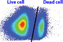
TMR/Cy3/Cy5 was introduced for the fluorescent label with peptides to evaluate each peptide’s cell-penetrating ability and intracellular distribution.
Cells (HeLa or Huh-7) were seeded on 24-well culture plates (40,000 cells/well) and incubated in 400 μL of DMEM containing 10% fetal bovine serum (FBS).
The medium was then replaced with fresh medium containing 10% FBS and Tetramethylrhodamine carboxylic acid (TMR)-labeled peptides.
The solution was added to each well at an appropriate concentration (e.g., 0.5 uM, 1 uM, 1.5 uM, 2 uM).
After 1, 2, 3, or 4 hours of incubation, the medium was removed, and cells were washed with ice-cold PBS and trypsin.
After adding medium containing 10% FBS, cells were centrifuged at 1600 rpm for 3 min at 4 °C. The cell pellets obtained were suspended in ice-cold PBS, centrifuged at 1600 rpm for 3 min at 4 °C, and then treated with Cell lysis buffer.
The fluorescence intensity of each lysate was measured using a spectrofluorometer. The amount of protein in each well was concomitantly determined using the BCA protein assay.
The results are presented as the mean and standard deviation from 3 samples.
Tumor antigens can be classified into two categories based on their expression pattern: tumor-specific antigens (TSA) and tumor-associated antigens (TAA).
Targeting tumor-associated antigens (TAAs) is a promising approach for cancer immunotherapy. Neoantigens are tumor-specific antigens originating from somatic mutations in cancer cells but not healthy tissues. So the TAAs are considered as ideal targets for novel immunotherapies. Antigens of three classes can induce tumor-specific T-cell responses.
1. Antigens derived from viral proteins: Viral proteins are produced inside the tumor cells. So the antigenic peptides can be detected by T cells.
2. Antigens derived from point mutations: Many CTL isolated from the tumors were found to recognize antigens that arise from point mutations in ubiquitously expressed genes. These mutations are passenger mutations, and the corresponding antigenic peptides are unique to the tumors in which they were identified.
3. Antigens encoded by cancer-germline genes: Cancer-germline genes are expressed in many cancer types and not in normal tissues except germline and trophoblastic cells. The tumor-specific pattern of expression results from the genome-wide demethylation in male germ cells.
A large number of antigenic peptides recognized by antitumor CTL have been identified. Candidate peptides can be synthesized and tested for HLA binding in vitro. The elution of antigenic peptides from MHC class I molecules immunopurified from the surface of tumor cells can be used to identify the antigens. TAAs can be targeted using peptide vaccines or by cellular approaches. The delivery of new peptide drugs might show great promise for future therapies.
Tumor-associated peptide antigens
LifeTein can customize a discovery and development path to fit your exact needs for peptide synthesis.
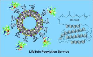
Polypeptides are used as new drug candidates to target specific disease symptoms. However, peptide drugs are rapidly degraded by proteolytic enzymes and neutralized by antibodies. Pegylation of polypeptide drugs improves their pharmacodynamic and pharmacokinetic profiles. Pegylating specifically can minimize the loss of biological activity and reduce immunogenicity. LifeTein offers peptide pegylation service and the PEG-modification of peptides through primary amines and sulfhydryl groups.
A significant limitation of the present PEGylated peptides is their heterogeneous nature because PEG is conjugated at many different nucleophilic amine residues. LifeTein’s approach to peptide PEGylation can address the fundamental issues of site-specific conjugation and high-efficiency conjugation. The click chemistry is widely used in the pegylation process.
The efficient ratio of 1:1 PEGylation of a peptide can be completed in 24 hours and purification of the PEG-protein conjugate in another three hours, without destroying their tertiary structure or abolishing their biological activity.
LifeTein’s improved technology is the use of branched structures, in contrast to the linear structures. Branched PEGs have increased molecular masses of up to 60 kDa or more, which is good at cloaking the attached polypeptide drug from the immune system and proteolytic enzymes.
Pegylation is the established method for improving the pharmacokinetics and pharmacodynamics of peptide pharmaceuticals.
New frontiers for the technology are now emerging for PEG-based hydrogels and PEG-modified liposomes, small-molecule modification, and the primary targets for pegylation of small-molecule drugs, oligonucleotides, lipids, cofactors, antibodies, saccharides, and nanoparticles.
Pegylation service from LifeTein
Manage Consent
To provide the best experiences, we use technologies like cookies to store and/or access device information. Consenting to these technologies will allow us to process data such as browsing behavior or unique IDs on this site. Not consenting or withdrawing consent, may adversely affect certain features and functions.
Functional
Always active
The technical storage or access is strictly necessary for the legitimate purpose of enabling the use of a specific service explicitly requested by the subscriber or user, or for the sole purpose of carrying out the transmission of a communication over an electronic communications network.
Preferences
The technical storage or access is necessary for the legitimate purpose of storing preferences that are not requested by the subscriber or user.
Statistics
The technical storage or access that is used exclusively for statistical purposes.The technical storage or access that is used exclusively for anonymous statistical purposes. Without a subpoena, voluntary compliance on the part of your Internet Service Provider, or additional records from a third party, information stored or retrieved for this purpose alone cannot usually be used to identify you.
Marketing
The technical storage or access is required to create user profiles to send advertising, or to track the user on a website or across several websites for similar marketing purposes.