Lipidation is a post-translational modification where a lipid molecule is covalently attached to a peptide or protein. This modification can significantly alter the peptide’s properties, including its solubility, stability, and cellular uptake. Understanding whether your peptide should be lipidated is crucial for optimizing its performance in various applications.
Key Takeaways
- Lipidation can alter peptide solubility and enhance stability.
- It can improve cellular uptake and membrane interaction.
- Consider the specific application and desired properties of your peptide.
Introduction to Peptide Lipidation
What are Lipidated Peptides?
Lipidation involves the attachment of lipid groups to peptides, which can include fatty acids, isoprenoids, or glycosylphosphatidylinositol (GPI) anchors. This modification can occur naturally or be introduced synthetically to enhance certain properties of the peptide.
Importance of Lipidated Peptides
Lipidation can significantly impact the biophysical properties of peptides. For instance, lipidated peptides often exhibit increased hydrophobicity, which can enhance their interaction with cell membranes and improve their bioavailability.
Benefits of Lipidating Peptides
Enhanced Solubility and Stability
Lipidation can improve the solubility of peptides in lipid environments, which is particularly beneficial for peptides intended for membrane-associated applications. Additionally, lipidated peptides often show increased stability against enzymatic degradation.
Improved Cellular Uptake
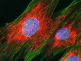
Lipidated peptides can more easily penetrate cell membranes, enhancing their cellular uptake and making them more effective in intracellular targeting. This is particularly useful for therapeutic peptides that need to reach intracellular targets.
Increased Membrane Interaction
The hydrophobic nature of lipidated peptides allows for better membrane interaction, which can be advantageous for peptides designed to disrupt or fuse with cell membranes.
Find Lipidation and more modifications here.
Considerations for Lipidation
Application-Specific Requirements
The decision to lipidate a peptide should be based on the specific application and the desired properties of the peptide. For example, peptides intended for therapeutic use may benefit from lipidation to enhance their bioavailability and stability.
Potential Drawbacks
While lipidation offers many benefits, it can also introduce challenges. Lipidated peptides may exhibit reduced solubility in aqueous environments and may require specialized formulation strategies.
Mechanisms of Lipidation
Types of Lipidated Peptides
Lipidation can occur through various mechanisms, each attaching different lipid groups to the peptide. Common types include:
- N-terminal myristoylation: Attachment of a myristoyl group to the N-terminal.
- S-palmitoylation: Addition of a palmitoyl group to cysteine residues.
- N-terminal stearylation: Attachment of a stearyl group to the N-terminal.

Synthetic Lipidation Techniques
Synthetic lipidation involves chemical methods to attach lipid groups to peptides. Techniques such as solid-phase peptide synthesis (SPPS) allow for precise control over the lipidation process, enabling the creation of peptides with specific properties.
Case Studies of Lipidated Peptides
Therapeutic Applications
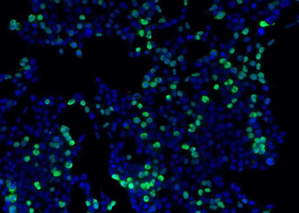
Lipidated peptides have shown promise in various therapeutic applications. For instance, lipidated antimicrobial peptides exhibit enhanced membrane-disruptive activity, making them effective against resistant bacterial strains.
Vaccine Development
In vaccine development, lipidated peptides can serve as potent adjuvants, enhancing the immune response. Lipidation can improve the delivery and presentation of antigens to the immune system, leading to stronger and more durable immunity.
Drug Delivery Systems
Lipidated peptides are also used in drug delivery systems to improve the targeting and release of therapeutic agents. By incorporating lipidated peptides into liposomes or nanoparticles, researchers can achieve more efficient delivery to specific tissues or cells.
Guidelines for Deciding on Lipidation
Assessing Peptide Properties
Before deciding to lipidate a peptide, assess its intrinsic properties such as solubility, stability, and target interaction. Lipidation may be beneficial if the peptide requires enhanced membrane interaction or cellular uptake.
Application-Specific Considerations
Consider the specific application of the peptide. For therapeutic peptides, lipidation can improve bioavailability and efficacy. For research applications, lipidation may facilitate cellular studies and membrane assays.
Potential Challenges
Be aware of potential challenges such as reduced aqueous solubility and the need for specialized formulation strategies. Balancing the benefits and drawbacks of lipidation is crucial for optimizing peptide performance.
Find our peptide synthesis services here.
Frequently Asked Questions
What are the benefits of lipidating my peptide?
Lipidation can alter peptide solubility and enhance stability, cellular uptake, and membrane interaction, making it beneficial for various applications, including therapeutics and drug delivery.
Are there any drawbacks to lipidation?
Yes, lipidation can reduce the peptide’s solubility in aqueous environments and may require specialized formulation strategies to maintain its effectiveness.
How do I decide if my peptide should be lipidated?
Consider the specific application and desired properties of your peptide. Assess its intrinsic properties and potential benefits of lipidation, such as improved bioavailability and stability.
Can lipidation be applied to any peptide?
While many peptides can be lipidated, the suitability depends on the peptide’s sequence and structure. Consulting with experts or using specialized services can help determine the best approach.