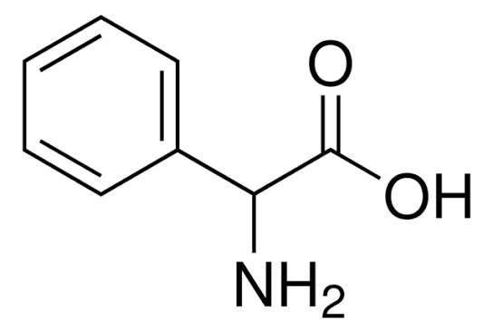
Phenylglycine (Phg) is a remarkable non-proteinogenic aromatic amino acid that has captured the attention of medicinal chemists and peptide scientists alike. Structurally, it resembles phenylalanine but lacks the methylene bridge (-CH₂-) between the amino acid backbone and the aromatic ring, resulting in a direct attachment of the phenyl group to the α-carbon. This subtle yet critical difference endows Phg with unique conformational properties and enhanced metabolic stability, making it an invaluable building block for the design of bioactive peptides and pharmaceuticals. However, the same structural features that make Phg so attractive also introduce significant synthetic challenges, particularly the risk of racemization during solid-phase peptide synthesis (SPPS). Understanding the properties, applications, and handling of this unusual amino acid is essential for researchers seeking to leverage its full potential.
Key Takeaways
- Phenylglycine is a non-proteinogenic amino acid characterized by a phenyl ring directly attached to the α-carbon, conferring unique conformational constraints and enhanced stability.
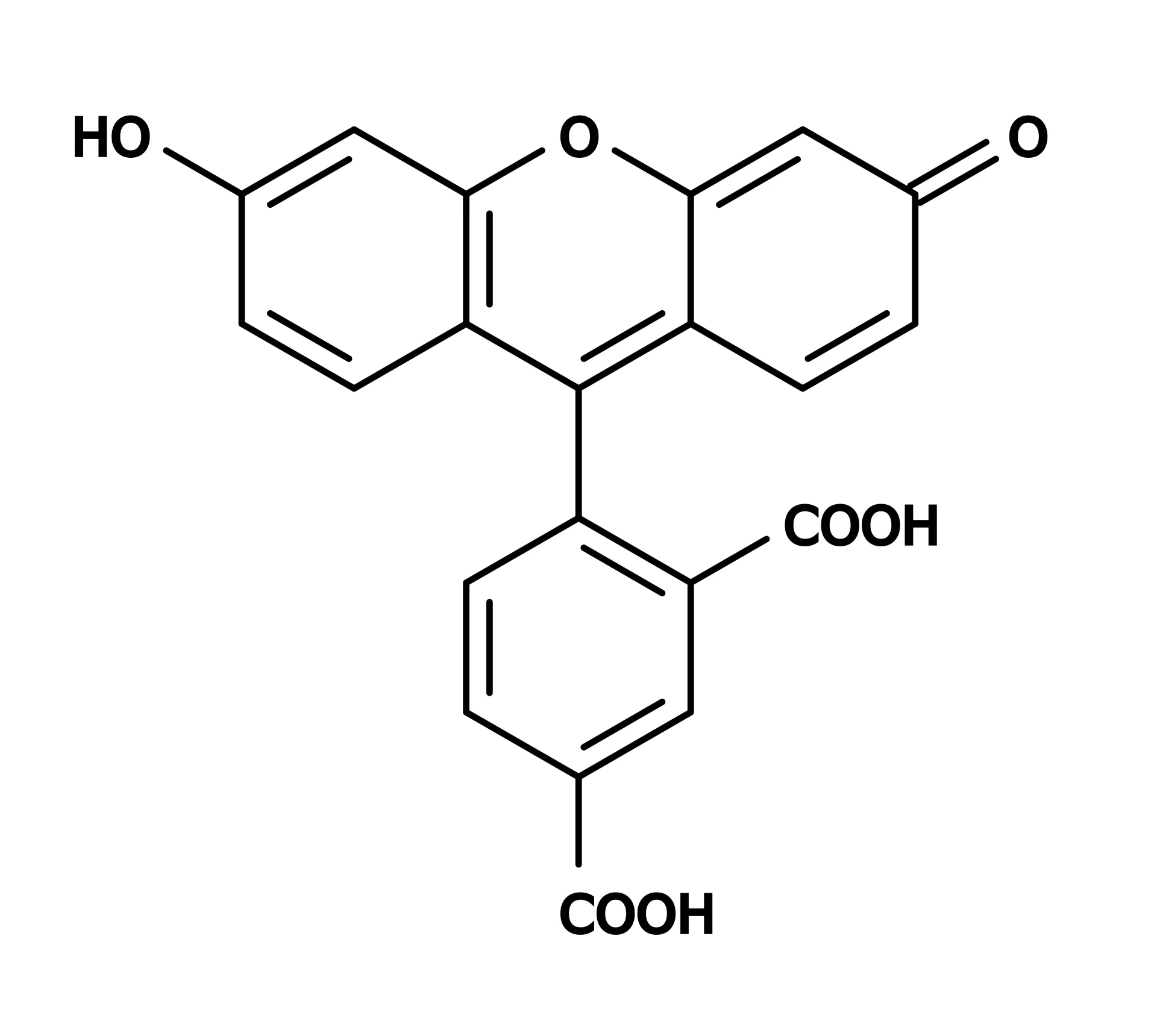
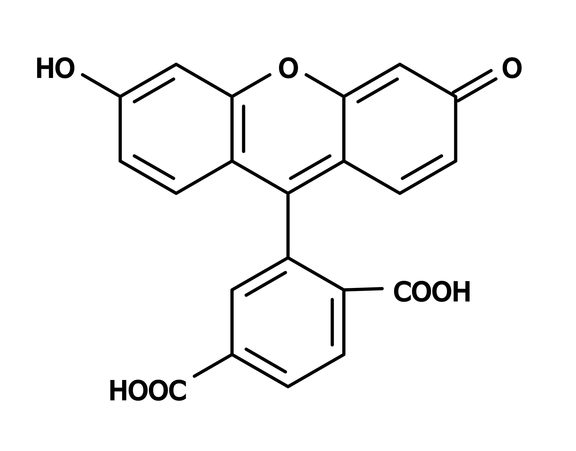
- Phg and its derivatives (e.g., 4-hydroxyphenylglycine, 3,5-dihydroxyphenylglycine) are crucial components of numerous peptide natural products, including glycopeptide antibiotics like vancomycin and streptogramins.
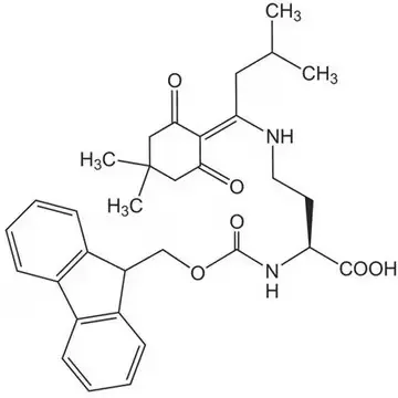
- The primary synthetic challenge is the high risk of racemization during Fmoc-SPPS, which can be mitigated by using specialized coupling reagents such as COMU or DEPBT with TMP or DMP.
- Peptides incorporating D-phenylglycine exhibit resistance to proteolytic degradation, extending their biological half-life.
- Phg-containing peptides have demonstrated promising biological activity against viral proteases (hepatitis C, dengue, West Nile Virus) and as antimicrobial agents.
- Custom synthesis of Phg-containing peptides is available through specialized providers equipped to handle the unique challenges of this residue.
Chemical Fundamentals of Phenylglycine
Structural Characteristics
Phenylglycine is formally defined as 2-amino-2-phenylacetic acid, with the molecular formula C₈H₉NO₂. Its defining structural feature is the direct attachment of the phenyl ring to the α-carbon of the amino acid backbone, which eliminates the benzylic methylene group present in phenylalanine. This structural distinction has profound conformational consequences: the Phg residue is significantly more constrained than phenylalanine, restricting the rotational freedom of the side chain and influencing the peptide backbone geometry.
Isomeric Forms and Chirality
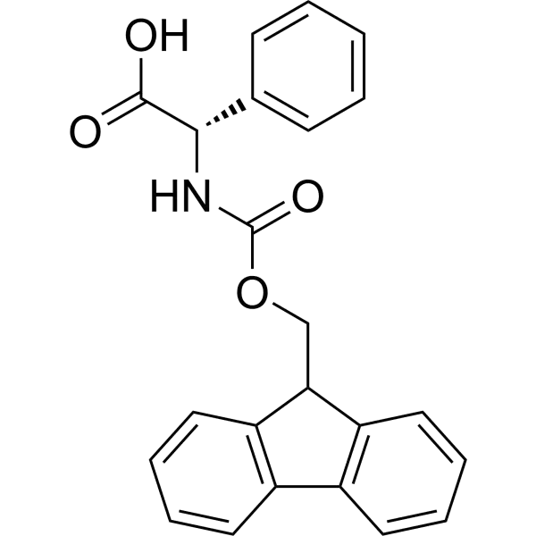
Phg exists as two enantiomers: L-phenylglycine and D-phenylglycine. Both forms are commercially available as Fmoc-protected building blocks for SPPS. Interestingly, the L-isomer is a rare natural product found in certain peptide antibiotics, while the D-isomer is widely used as a side-chain building block for semi-synthetic penicillins and cephalosporins, including ampicillin and cephalexin. The choice of enantiomer profoundly influences both the biological activity and the conformational behavior of the resulting peptides.
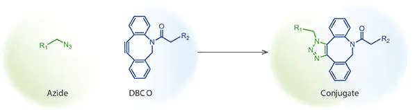
Find out more about peptide synthesis here.
Biological Significance and Natural Occurrence
Occurrence in Peptide Natural Products
Phg-type amino acids are widely distributed in nature as components of numerous biologically active peptide natural products. The most important representatives are phenylglycine (Phg), 4-hydroxyphenylglycine (Hpg), and 3,5-dihydroxyphenylglycine (Dpg). These unusual residues are essential building blocks of:
- Glycopeptide antibiotics: Vancomycin and related compounds rely on Phg derivatives for their rigid three-dimensional structure and antibiotic activity.
- Streptogramins: Antibiotics such as pristinamycin I and virginiamycin S contain L-phenylglycine residues.
- Other bioactive peptides: Phg is also found in the bicyclic peptide antibiotic dityromycin and various linear and cyclic peptides with diverse biological activities.
Role in Medicinal Chemistry
The significance of phenylglycine for medicinal chemistry has drawn the attention of many research groups and pharmaceutical companies. Beyond its natural occurrence, Phg-containing peptides have shown promising biological activity against viral targets, including inhibitors of hepatitis C, dengue, and West Nile Virus proteases. Furthermore, D-phenylglycine is actively utilized to modulate the pharmacokinetics of peptide drugs; because D-amino acids are mirror images of naturally occurring L-amino acids, they are inherently resistant to endogenous proteases, thereby extending the biological half-life of therapeutic agents.
Conformational Properties
Unique Structural Constraints
The direct attachment of the phenyl ring to the α-carbon imposes significant conformational restrictions on Phg-containing peptides. Quantum mechanical and molecular dynamics studies have revealed that the conformational states of Phg dipeptides are stabilized by non-covalent interactions, including carbonyl-carbonyl interactions and carbonyl-π (aromatic ring) interactions. In simulation studies, the Φ, ψ values for the R-form of Phg correspond to the inverse β/inverse collagen region, while the S-form adopts conformations in the β/collagen region. This conformational behavior is of great value for designing bioactive peptides that require a single, well-defined conformation for target recognition.
Synthesis and Handling Challenges
The Racemization Problem
Despite its utility, phenylglycine is often considered a troublesome residue in peptide synthesis. The primary challenge is the high risk of racemization (epimerization) during Fmoc-based SPPS. The acidic α-proton of Phg is particularly labile due to the electron-withdrawing effect of the directly attached phenyl ring, making it susceptible to base-catalyzed racemization during coupling steps.
Strategies for Minimizing Racemization
Research has demonstrated that the base-catalyzed coupling of Fmoc-Phg is the critical step for racemization. However, racemization can be reduced to a negligible level by employing specific coupling reagents and conditions. The recommended approach utilizes:
- COMU or DEPBT as coupling reagents
- TMP (2,4,6-trimethylpyridine) or DMP (2,6-dimethylpyridine) as bases
These reagents minimize epimerization while maintaining high coupling efficiency. Importantly, resin-bound Phg-containing peptides are remarkably resistant to epimerization during extended incubation under basic conditions, and the free peptides are stable in buffer solutions used for biological assays.
Commercial Availability
For researchers incorporating Phg into custom peptides, Fmoc-protected building blocks are commercially available:
- Fmoc-Phg-OH for L-phenylglycine
- Fmoc-D-Phg-OH for D-phenylglycine
Specialized providers such as LifeTein offer custom synthesis of Phg-containing peptides, with expertise in handling the unique challenges of this residue, including optimized coupling protocols to minimize racemization and ensure high purity.
Find out about high-speed RUSH synthesis.
Frequently Asked Questions (FAQ)
What is the difference between phenylglycine and phenylalanine?
Phenylglycine (Phg) has the phenyl ring directly attached to the α-carbon (2-amino-2-phenylacetic acid), whereas phenylalanine (Phe) has a methylene bridge (-CH₂-) between the α-carbon and the phenyl ring (2-amino-3-phenylpropionic acid). This structural difference makes Phg significantly more conformationally constrained and provides enhanced metabolic stability.
Why is phenylglycine considered a “difficult” amino acid in peptide synthesis?
Phenylglycine is prone to racemization (epimerization) during Fmoc-based SPPS because its α-proton is highly acidic due to the electron-withdrawing effect of the directly attached phenyl ring. This can lead to loss of stereochemical integrity if not carefully controlled.
How can racemization of phenylglycine be prevented during synthesis?
Racemization can be minimized by using COMU or DEPBT as coupling reagents in combination with TMP or DMP as bases. These conditions have been shown to reduce racemization to a negligible level while maintaining high coupling efficiency.
What are the main applications of phenylglycine-containing peptides?
Phg-containing peptides have diverse applications, including:
- Antimicrobial agents: As components of glycopeptide antibiotics (vancomycin) and streptogramins
- Antiviral agents: Inhibitors of hepatitis C, dengue, and West Nile Virus proteases
- Protease-resistant therapeutics: D-phenylglycine imparts resistance to proteolytic degradation, extending biological half-life
Can D-phenylglycine be incorporated into peptides?
Yes. Fmoc-D-Phg-OH is commercially available as a standard building block for Fmoc SPPS. Incorporation of D-phenylglycine is a valuable strategy for enhancing proteolytic stability and modulating the pharmacokinetics of peptide drugs.
References
Liang, C., Behnam, M. A. M., Sundermann, T. R., & Klein, C. D. (2017). Phenylglycine racemization in Fmoc-based solid-phase peptide synthesis: Stereochemical stability is achieved by choice of reaction conditions. Tetrahedron Letters, 58(24), 2325–2329. https://doi.org/10.1016/j.tetlet.2017.04.047
Nandel, F. S., & Shafique, M. (2014). Conformational behavior of phenylglycines and hydroxyphenylglycines and non-planarity of phenyl rings. Indian journal of biochemistry & biophysics, 51(5), 350–357.
Voitsekhovskaia, I., Ho, Y. T. C., Klatt, C., Müller, A., Machell, D. L., Tan, Y. J., Triesman, M., Bingel, M., Schittenhelm, R. B., Tailhades, J., Kulik, A., Maier, M. E., Otting, G., Wohlleben, W., Schneider, T., Cryle, M., & Stegmann, E. (2024). Altering glycopeptide antibiotic biosynthesis through mutasynthesis allows incorporation of fluorinated phenylglycine residues. RSC Chemical Biology, 5(10), 1017–1034. https://doi.org/10.1039/d4cb00140k
Al Toma, R. S., Brieke, C., Cryle, M. J., & Süssmuth, R. D. (2015). Structural aspects of phenylglycines, their biosynthesis and occurrence in peptide natural products. Natural Product Reports, 32(8), 1207–1235. https://doi.org/10.1039/c5np00025d