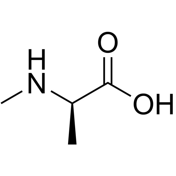
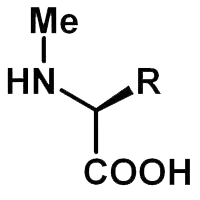
FLAG: The Epitope Tag Peptide

FLAG

The FLAG peptide, also known as DYKDDDDK, is a short, hydrophilic, and highly charged peptide sequence. It is extensively utilized in the field of molecular biology for the detection and purification of proteins. Delve into the significance of the FLAG peptide, its applications, and its advantages in scientific research.

Key Takeaways

  • FLAG peptide is a widely used epitope tag in molecular biology.
  • It facilitates protein purification and detection.
  • The DYKDDDDK sequence is highly specific and efficient.

Structure and Properties of FLAG Peptide

Amino Acid Sequence

The FLAG peptide consists of eight amino acids: Asp-Tyr-Lys-Asp-Asp-Asp-Asp-Lys. This sequence is designed to be highly specific and efficient in binding to antibodies, making it an ideal choice for various biochemical applications.

Find our FLAG products here.

Hydrophilicity and Charge

The peptide is highly hydrophilic and carries a significant negative charge due to the presence of multiple aspartic acid residues. These properties enhance its solubility and interaction with other molecules, facilitating its use in different experimental conditions.

Applications of FLAG Peptide

Protein Purification

One of the primary applications of the FLAG peptide is in protein purification. By fusing the FLAG tag to a target protein, researchers can easily isolate and purify the protein using affinity chromatography techniques. The FLAG tag binds specifically to anti-FLAG antibodies, allowing for efficient separation from other cellular components.

Protein Detection

The FLAG peptide is also widely used for protein detection in various assays, including Western blottingimmunoprecipitation, and immunofluorescence. The high specificity of the FLAG tag ensures accurate detection of the target protein, even in complex mixtures.

Advantages of Using FLAG Peptide

High Specificity

The FLAG peptide’s sequence is highly specific, reducing the likelihood of cross-reactivity with other proteins. This specificity is crucial for obtaining accurate and reliable results in both purification and detection assays.

Versatility

The FLAG tag can be used in a variety of experimental setups, making it a versatile tool in molecular biology. Its compatibility with different techniques and conditions enhances its utility in diverse research applications.

Ease of Use

The FLAG peptide is easy to use and can be readily incorporated into experimental protocols. Its well-characterized properties and availability of high-quality antibodies further simplify its application in research.

Find more peptide synthesis here.

The FLAG Peptide

FLAG

Frequently Asked Questions

What is the FLAG peptide sequence?

  • The FLAG peptide sequence is Asp-Tyr-Lys-Asp-Asp-Asp-Asp-Lys (DYKDDDDK).

How does the FLAG tag facilitate protein purification?

  • The FLAG tag binds specifically to anti-FLAG antibodies, allowing for efficient isolation of the tagged protein using affinity chromatography techniques.

Can the FLAG peptide be used in different experimental conditions?

  • Yes, the FLAG peptide is highly versatile and can be used in various experimental setups, including different buffers and conditions.

What are the advantages of using the FLAG peptide over other tags?

  • The FLAG peptide offers high specificity, ease of use, and compatibility with multiple detection and purification techniques, making it a preferred choice in molecular biology research.

PADRE Peptide: Advancing Immunotherapy and Vaccines

PADRE

The PADRE peptide (Pan-DR epitope) is a synthetic peptide designed to bind to a wide range of human MHC class II molecules. This characteristic makes it a universal helper T-cell epitope, which is crucial in enhancing immune responses. The PADRE peptide is particularly valuable in the field of vaccine development due to its ability to stimulate robust T-cell responses.

Mechanism of Action

Key Takeaways:

  • PADRE peptide is a universal helper T-cell epitope.
  • It enhances immune responses in vaccine development.
  • Applications include cancer immunotherapy and infectious disease vaccines.

Introduction to PADRE Peptide

Binding to MHC Class II Molecules

The PADRE peptide’s primary function is to bind to MHC class II molecules, which are essential for presenting antigens to helper T-cells. This binding facilitates the activation of T-cells, which in turn, enhances the overall immune response. The broad binding affinity of PADRE to various MHC class II molecules makes it a versatile tool in immunotherapy.

Enhancing Immune Responses

By acting as a universal helper T-cell epitope, PADRE can significantly boost the immune response to various antigens. This enhancement is particularly beneficial in cancer immunotherapy and infectious disease vaccines, where a strong and targeted immune response is crucial for efficacy.

PADRE Applications in Cancer Immunotherapy

Targeting Tumor Antigens

In cancer immunotherapy, the PADRE peptide is used to enhance the immune system’s ability to recognize and attack tumor cells. By incorporating PADRE into peptide-based vaccines, researchers can improve the presentation of tumor antigens to the immune system, leading to a more effective anti-tumor response.

Discover more peptide synthesis here.

Clinical Trials and Research

Several clinical trials have explored the use of PADRE in cancer vaccines. For instance, vaccines targeting Mucin 1 (MUC1), a glycoprotein overexpressed in many cancers, have shown promising results when combined with PADRE. These vaccines have demonstrated the ability to elicit strong immune responses, including the production of antibodies against cancer-specific antigens.

PADRE Applications in Infectious Disease Vaccines

Enhancing Vaccine Efficacy

The PADRE peptide is also employed in the development of vaccines for infectious diseases. By incorporating PADRE into vaccine formulations, researchers can enhance the immune response to various pathogens. This approach has been particularly useful in developing vaccines for diseases with high mutation rates, such as influenza and HIV.

Case Studies and Examples

One notable example is the development of a peptide-based vaccine for the H1N1 influenza virus. Researchers have identified a killer decapeptide (KP) with potent action against the virus. When combined with PADRE, this vaccine has shown improved efficacy in reducing viral levels and improving survival rates in animal models.

Applications in Autoimmune Diseases

Modulating Immune Responses

The PADRE peptide has shown potential in the treatment of autoimmune diseases by modulating immune responses. In conditions such as rheumatoid arthritis and multiple sclerosis, the immune system mistakenly attacks the body’s own tissues. By incorporating PADRE into therapeutic strategies, researchers aim to redirect the immune response, reducing inflammation and tissue damage.

Preclinical and Clinical Studies

Preclinical studies have demonstrated that PADRE can induce regulatory T-cells (Tregs), which play a crucial role in maintaining immune tolerance. These findings have paved the way for clinical trials exploring PADRE-based therapies for autoimmune diseases. Early results indicate that PADRE can help restore immune balance, offering a promising avenue for treatment.

Applications in Allergy Treatments

Reducing Allergic Reactions

In allergy treatments, the PADRE peptide is used to reduce hypersensitivity reactions. By enhancing the immune system’s ability to tolerate allergens, PADRE can help mitigate symptoms associated with allergic conditions such as asthma and food allergies.

Immunotherapy Approaches

Immunotherapy approaches incorporating PADRE have shown efficacy in desensitizing patients to specific allergens. For example, PADRE-based vaccines targeting peanut allergies have demonstrated the ability to reduce allergic reactions in clinical trials. These vaccines work by gradually exposing the immune system to the allergen in a controlled manner, promoting tolerance.

Find the PADRE Peptide here.

PADRE

Future Directions and Research

Expanding Therapeutic Applications

Ongoing research aims to expand the therapeutic applications of the PADRE peptide. Scientists are exploring its potential in areas such as transplantation medicine, where PADRE could help prevent organ rejection by modulating the immune response. Additionally, PADRE is being investigated for its role in enhancing the efficacy of DNA vaccines and mRNA vaccines, which have gained prominence in recent years.

Innovative Delivery Systems

Innovative delivery systems are being developed to improve the stability and efficacy of PADRE-based therapies. These include nanoparticle-based delivery and liposomal formulations, which can enhance the bioavailability and targeted delivery of PADRE to specific tissues.

Frequently Asked Questions

What is the primary function of the PADRE peptide? 

  • The primary function of the PADRE peptide is to bind to MHC class II molecules, enhancing the activation of helper T-cells and boosting immune responses.

How is PADRE used in cancer immunotherapy? 

  • In cancer immunotherapy, PADRE is incorporated into peptide-based vaccines to improve the presentation of tumor antigens to the immune system, leading to a more effective anti-tumor response.

Can PADRE be used in the treatment of autoimmune diseases? 

  • Yes, PADRE has shown potential in modulating immune responses in autoimmune diseases, helping to reduce inflammation and tissue damage.

What are some examples of PADRE’s applications in allergy treatments? 

  • PADRE is used in immunotherapy approaches to reduce allergic reactions, such as in vaccines targeting peanut allergies, which promote immune tolerance to the allergen.

What future research directions are being explored for PADRE? 

  • Future research is exploring PADRE’s potential in transplantation medicine, DNA and mRNA vaccines, and innovative delivery systems like nanoparticle-based and liposomal formulations.

Fluorescent Labeling with Alexa Fluor 647

Alexa Fluor 647

Fluorescent peptides play a crucial role in biological research, enabling the visualization and tracking of molecules and processes. Among the various fluorescent dyes available, Alexa Fluor 647 stands out due to its bright and photostable properties. Delve into fluorescent peptide labeling with Alexa Dye 647, exploring its applications, advantages, and considerations.

Introduction to Alexa Fluor 647

Alexa Fluor 647 is a succinimidyl ester dye that efficiently reacts with primary amines of proteins, forming stable dye–protein conjugates. Here are some key points about Alexa Fluor 647:

  • Bright and Photostable: Alexa Fluor 647 emits light at approximately 668 nm, making it ideal for multicolor applications. Its wide spectral separation from other red fluorophores ensures minimal interference.
  • pH Insensitive: The fluorescent signal remains consistent between pH 4 and 10, allowing reliable imaging across different cellular environments.
  • Resistant to Quenching: Unlike Cy™5 dye, Alexa Fluor 647 maintains fluorescence even at high degrees of substitution.

Find more Fluorescents here.

Applications of Alexa Fluor 647-Labeled Peptides

  1. Fluorescence Microscopy: Alexa Fluor 647-labeled peptides serve as excellent probes for visualizing cellular structures, protein localization, and dynamic processes.
  2. Protein-Protein Interactions: Researchers use these labeled peptides to study interactions between proteins, shedding light on signaling pathways and molecular networks.
  3. Enzymatic Activity Monitoring: By attaching Alexa Fluor 647 to specific peptide substrates, enzymatic activities can be tracked in real-time.
  4. FRET Assays: Fluorescence Resonance Energy Transfer (FRET) studies benefit from Alexa Fluor 647 as both donor and acceptor fluorophores.
Alexa Fluor 647

Considerations and Best Practices

  • Buffer Choice: To achieve optimal labeling efficiency, ensure the purified protein is in a buffer free of ammonium ions or primary amines.
  • Protein Purity: Impure proteins, such as antibodies in crude serum, may not label well.
  • Storage: Store Alexa Fluor 647 at ≤–20°C and protect it from light. Components B-D should be stored at 2–6°C.
  • Fluorescent Dye Selection: Based on your specific experimental needs, consider other fluorescent dyes (e.g., FITC, Cy3, Cy5).

Practical Tips for Successful Peptide Labeling with Alexa Fluor 647

Conjugation Strategies

  1. Direct Labeling: In direct labeling, the dye is directly attached to the peptide. This method is straightforward but requires careful optimization of reactant ratios and reaction conditions.
  2. Two-Step Labeling: Here, the peptide is first modified with a reactive group (e.g., Cys-Maleimide, Lys(N3), NHS ester) and then conjugated to Alexa Fluor 647. This approach allows better control over labeling efficiency.

Find more about Peptide Synthesis here.

Frequently Asked Questions

Q1: Can I use Alexa Fluor 647 for live-cell imaging?

A: Yes! Alexa Fluor 647 is suitable for live-cell imaging due to its photostability and minimal photobleaching. However, cell permeability and potential cytotoxicity should be considered.

A: Store labeled peptides at –20°C in the dark. Components B-D should be stored at 2–6°C.

Q3: Can I multiplex Alexa Fluor 647 with other fluorophores?

A: Absolutely! Alexa Fluor 647 pairs well with other dyes like FITC, Cy3, and Cy5. Optimize spectral overlap for multicolor experiments.

Click Chemistry in Peptide Synthesis

Click Chemistry

Click chemistry, a groundbreaking technique recognized by the 2022 Nobel Prize in Chemistry, has revolutionized peptide synthesis. Developed by Carolyn R. Bertozzi, Morten Meldal, and K. Barry Sharpless, this method enables efficient and precise conjugation of peptides with other biomolecules. At the forefront of this innovation, LifeTein leverages click chemistry to create peptide-drug conjugates with remarkable applications.

Key Takeaways

  • Click Chemistry Defined: Click chemistry involves the selective and irreversible coupling of two molecular components. In peptide synthesis, this technique allows for precise and efficient conjugation.
  • Cu (I)-Catalyzed Azide-Alkyne Click Chemistry (CuAAC): LifeTein employs CuAAC to link peptides with azide and alkyne functional groups. This robust reaction enables the creation of peptide-drug conjugates.
  • Strain-Promoted Azide-Alkyne Click Chemistry (SPAAC): SPAAC, a copper-free variant, facilitates bioconjugation without the need for metal catalysts.
  • Tetrazine-Alkene Ligation: LifeTein also utilizes tetrazine-alkene ligation, a powerful click chemistry approach, for peptide-drug conjugation.

Applications

  1. Peptide-Drug Conjugation: LifeTein’s expertise extends to developing peptide-drug and antibody-drug conjugates. Successful conjugations involve drugs like MMAE, Panobinostat, Tazmetostat, and FK506.
  2. Site-Specific PEGylation: Using maleimide or click chemistry, LifeTein achieves site-specific PEGylation of peptides. These PEGylated peptides find applications in various fields.
  3. Photo Crosslinking Peptides: Incorporating photo-labile amino acids, such as p-benzoyl-L-phenylalanine (Bpa), LifeTein enables downstream click chemistry. Functionalized alkynes, fluorophores, and other groups can be efficiently linked to peptides.

Explore LifeTein’s Custom Synthesis Services

Click Chemistry

Mechanisms of Click Chemistry

Cu (I)-Catalyzed Azide-Alkyne Click Chemistry (CuAAC)

CuAAC, also known as the Huisgen reaction, remains a cornerstone in peptide synthesis. Here’s how it works:

  1. Azide and Alkyne Functionalization: Peptides are modified with azide (–N₃) and alkyne (–C≡CH) groups.
  2. Copper(I) Catalyst: In the presence of a copper(I) catalyst, the azide and alkyne react to form a stable triazole linkage.
  3. High Yield and Selectivity: CuAAC offers excellent yield and selectivity, making it ideal for peptide conjugation.

Strain-Promoted Azide-Alkyne Click Chemistry (SPAAC)

SPAAC, a copper-free variant, avoids the need for metal catalysts. Key features:

  1. Strain-Driven Reaction: The reaction relies on strained cyclooctynes (e.g., DBCO) and azides. No copper required!
  2. Bioorthogonal: SPAAC occurs selectively in biological environments without interfering with native biomolecules.

Tetrazine-Alkene Ligation

LifeTein also employs tetrazine-alkene ligation for peptide-drug conjugation:

  1. Tetrazine and Alkene: Peptides are functionalized with tetrazine and alkene groups.
  2. Rapid Reaction: Tetrazine reacts with alkene (e.g., trans-cyclooctene) in a bioorthogonal, rapid ligation.

FAQ

Q: What advantages does click chemistry offer in peptide synthesis?

A: Click chemistry provides precise, efficient conjugation, enabling the creation of peptide-drug conjugates and other bioconjugates.

Q: Which drugs are successfully conjugated by LifeTein?

A: LifeTein excels in conjugating MMAE, Panobinostat, Tazmetostat, and FK506 to peptides.

Q: Can LifeTein perform site-specific PEGylation of peptides?

A: Yes! Using maleimide or click chemistry, LifeTein achieves site-specific PEGylation for versatile applications.


Peptide Conjugation Magnetic Beads

Magnetic Beads

Peptide conjugation magnetic beads are powerful tools in biotechnology and molecular biology. They facilitate the covalent attachment of peptides to a magnetic core, enabling efficient separation and purification processes.

Key Takeaways

  • Efficient separation: Magnetic beads allow for quick and easy separation of target molecules.
  • Versatile applications: Used in protein purification, immunoprecipitation, and diagnostic assays.
  • High binding capacity: Capable of binding significant amounts of peptides or proteins.

Introduction to Peptide Conjugation Magnetic Beads

What Are Magnetic Beads?

Magnetic beads are small, superparamagnetic particles that can be manipulated using an external magnetic field. These beads are typically coated with functional groups that allow for the covalent attachment of biomolecules such as peptides, proteins, and nucleic acids.

Importance of Peptide Conjugation

Peptide conjugation to magnetic beads enhances their utility in various applications. By attaching peptides to the beads, researchers can target specific molecules, enabling selective binding and separation. This is particularly useful in protein purification, immunoprecipitation, and diagnostic assays.

Types of Peptide Conjugation Magnetic Beads

Amine-Activated Magnetic Beads

Amine-activated magnetic beads are coated with primary amine groups, which can react with carboxyl or aldehyde groups on peptides. This type of bead is ideal for covalent coupling under mild conditions, preserving the activity of the peptides.

Thiol-Activated Magnetic Beads

Thiol-activated magnetic beads are functionalized with maleimide groups, which react with thiol groups on peptides. This specific interaction ensures a stable and efficient conjugation process, making these beads suitable for various bioseparation applications.

Applications of Peptide Conjugation Magnetic Beads

Protein Purification

One of the primary applications of peptide conjugation magnetic beads is in protein purification. By attaching specific peptides to the beads, researchers can selectively bind and isolate target proteins from complex mixtures. This method is highly efficient and can be scaled up for large-volume applications.

Immunoprecipitation

Immunoprecipitation is another critical application where peptide conjugation magnetic beads are used. In this technique, antibodies are conjugated to the beads, allowing for the selective capture of antigens from a sample. The magnetic properties of the beads facilitate easy separation and purification of the antigen-antibody complexes.

Diagnostic Assays

In diagnostic assays, peptide conjugation magnetic beads are used to detect specific biomarkers. The beads can be functionalized with peptides that bind to target molecules, enabling sensitive and specific detection in various diagnostic platforms.

Magnetic Beads

Preparation of Magnetic Beads

Washing and Buffer Exchange

Before conjugation, magnetic beads must be thoroughly washed and equilibrated in the appropriate buffer. This step ensures the removal of any preservatives or contaminants that might interfere with the conjugation process. Typically, beads are washed with a buffer such as phosphate-buffered saline (PBS) or sodium phosphate buffer.

Conjugation Process

Amine-Activated Beads

For amine-activated beads, the conjugation process involves the reaction of the primary amine groups on the beads with carboxyl or aldehyde groups on the peptides. This reaction is often facilitated by a coupling agent such as EDC (1-ethyl-3-(3-dimethylaminopropyl) carbodiimide)1. The reaction conditions are usually mild, preserving the biological activity of the peptides.

Thiol-Activated Beads

Thiol-activated beads utilize maleimide groups to react with thiol groups on peptides. This reaction is highly specific and efficient, forming a stable thioether bond. The conjugation is typically performed at physiological pH and room temperature, ensuring high yields and minimal peptide degradation.

Post-Conjugation Steps

Blocking and Washing

After conjugation, any remaining reactive groups on the beads must be blocked to prevent non-specific binding. Common blocking agents include bovine serum albumin (BSA) or ethanolamine. The beads are then washed to remove unbound peptides and other reaction by-products.

Storage Conditions

Proper storage of peptide-conjugated magnetic beads is crucial for maintaining their functionality. The beads should be stored at 2-8°C in a buffer containing a preservative such as sodium azide to prevent microbial growth.

Advanced Applications

High-Throughput Screening

Peptide conjugation magnetic beads are increasingly used in high-throughput screening (HTS) applications. Their ability to selectively bind target molecules makes them ideal for screening large libraries of compounds or peptides. This application is particularly valuable in drug discovery and biomarker identification.

Cell Separation

In addition to protein purification, peptide-conjugated magnetic beads are also used for cell separation. By attaching cell-specific peptides to the beads, researchers can isolate specific cell types from heterogeneous populations. This technique is widely used in immunology and stem cell research.

Proteomics

Proteomics, the large-scale study of proteins, benefits significantly from the use of peptide-conjugated magnetic beads. These beads facilitate the enrichment and identification of low-abundance proteins, enhancing the sensitivity and accuracy of proteomic analyses.

Frequently Asked Questions (FAQ)

What are the advantages of using peptide conjugation magnetic beads?

  • High specificity: Enables selective binding and separation of target molecules.
  • Versatility: Suitable for a wide range of applications, including protein purification, immunoprecipitation, and diagnostic assays.
  • Efficiency: Facilitates rapid and efficient separation processes.

How do I choose between amine-activated and thiol-activated beads?

The choice depends on the functional groups present on your peptides. Amine-activated beads are suitable for peptides with carboxyl or aldehyde groups, while thiol-activated beads are ideal for peptides with thiol groups.

What are the storage conditions for peptide-conjugated magnetic beads?

Peptide-conjugated magnetic beads should be stored at 2-8°C in a buffer containing a preservative such as sodium azide to maintain their stability and prevent microbial contamination.

Can peptide-conjugated magnetic beads be reused?

While it is possible to reuse peptide-conjugated magnetic beads, their binding capacity may decrease with each use. It is essential to follow proper washing and storage protocols to maximize their lifespan.

Fluorescent Labelling with Texas Red

Fluorescent labeling is a powerful technique used in cell biology and microscopy to visualize specific molecules within cells. Among the various fluorophores available, Texas Red stands out as a bright red-fluorescent dye commonly used for cellular imaging applications. It holds many vital applications in biological research.

Texas Red: Properties and Applications

  • Bright Fluorescence: Texas Red emits a vibrant red fluorescence when excited by laser lines at 561 or 594 nm. Its brightness makes it ideal for detecting weakly expressed antigens or proteins in biological samples.
  • Conjugation to Antibodies and Peptides: Researchers often conjugate Texas Red to antibodies or peptides. When these labeled molecules bind to specific targets (such as antigens), they reveal the location of those targets within cells.
  • Photostability: Texas Red exhibits good photostability in buffer and antifade conditions, allowing for reliable imaging over extended periods.
  • Alternative: Alexa Fluor 594: For even brighter and more photostable conjugates, consider using Invitrogen Alexa Fluor 594. It shares spectral properties with Texas Red but offers improved solubility and sensitivity.

Find more about Peptide Synthesis here.

Key Takeaways

  • Texas Red is a red-fluorescent dye commonly used for cellular imaging.
  • It is conjugated to antibodies and proteins to visualize specific targets within cells.
  • Researchers can choose between Texas Red and Alexa Fluor 594 based on their specific imaging needs.

Intracellular Localization Studies

Texas Red-labeled antibodies and peptides have been instrumental in studying the localization of specific molecules within cells. By targeting specific antigens or proteins, researchers can visualize their distribution in various cellular compartments:

Nucleus

  • Texas Red-conjugated antibodies against nuclear proteins (e.g., histones) allow precise visualization of the nucleus. This aids in understanding chromatin organization and gene expression.

Cytoskeleton

  • Texas Red-labeled phalloidin binds to actin filaments, revealing the intricate cytoskeletal network. Researchers use this to study cell motility, shape changes, and intracellular transport.

Membrane Proteins

  • Texas Red-labeled antibodies against membrane proteins (e.g., receptors) help identify their presence on the cell surface. This is crucial for signaling studies and drug development.

Multicolor Imaging

Texas Red is often used in multicolor experiments alongside other fluorophores. Combining it with green (e.g., FITC) or blue (e.g., DAPI) fluorophores allows simultaneous visualization of multiple targets within the same sample.

Find more Fluorescents here.

Texas Red

Frequently Asked Questions

  • What is Texas Red?
  • Texas Red is a red-fluorescent dye commonly used in cell biology and microscopy. It emits bright red fluorescence when excited by specific laser lines, making it ideal for visualizing specific molecules within cells.
  • How is Texas Red used in research?
  • Intracellular Localization Studies: Texas Red-labeled antibodies and proteins help researchers study the distribution of specific molecules within cellular compartments.
  • Can Texas Red be used alongside other dyes?
  • Yes, Texas Red can be used in multicolor experiments alongside other fluorophores. Combining it with different colors allows researchers to study multiple targets simultaneously.

Phosphorylated Peptides: A Key Figure in Biological Processes

Phosphorylated peptides

Phosphorylated peptides are a result of a widespread post-translational modification that occurs as a result of esterification of amino acid side chains in peptides. This process involves the addition of a strongly negatively charged phosphate group, thereby altering the protein’s conformation, activity, and ability to interact with other molecules.

Key Takeaways:

  • Phosphorylated peptides are a result of a post-translational modification.
  • The process involves the addition of a phosphate group to amino acid side chains in peptides.
  • This modification alters the protein’s conformation, activity, and ability to interact with other molecules.
  • Phosphorylated peptides play an important role in the regulation of many biological processes.

The Importance of Phosphorylated Peptides

Role in Biological Mechanisms

Phosphorylated peptides play an important role in the regulation of many biological processes, such as signaling, gene expression, and cell division. The mechanism of peptide phosphorylation is that the process of transferring the phosphoric acid group of GTP or ATP γ-position to the protein amino acid residue on the basis of kinase catalysis. This occurs mainly on the hydroxyl groups of serine, tyrosine and threonine residue side chains2. It plays an important role in signaling between cells.

Impact on Protein Function

Protein phosphorylation often activates (or deactivates) many enzymes. Phosphorylation is essential to the processes of both anaerobic and aerobic respiration, which involve the production of adenosine triphosphate (ATP), the “high-energy” exchange medium in the cell. During aerobic respiration, ATP is synthesized in the mitochondrion by addition of a third phosphate group to adenosine diphosphate (ADP) in a process referred to as oxidative phosphorylation.

Challenges in Studying Phosphorylated Peptides

While protein regions that do not contain post-translational modifications (PTMs) can be rather simply mimicked using peptide libraries, heavily phosphorylated regions are much harder to study using the same tools. The differences between the syntheses of simple mono-, di- and tri-phosphopeptides and the synthesis of multiphosphopeptides are dramatic. Synthesis of multiphosphopeptides requires the insertion of several phosphate groups simultaneously or sequentially into various positions on the peptide in the presence of many other potential modification sites.

Find our list of modifications here.

Phosphorylated peptides


Synthetic Strategies for Phosphorylated Peptides

Traditional Methods

Traditional methods for the synthesis of phosphorylated peptides involve the use of phosphoramidite or phosphotriester chemistry. These methods, while effective, can be time-consuming and require the use of harsh reaction conditions. Additionally, these methods often result in the formation of by-products that can complicate the purification process.

Modern Techniques

Modern techniques for the synthesis of phosphorylated peptides have focused on improving the efficiency and selectivity of the phosphorylation process. One such technique involves the use of solid-phase peptide synthesis (SPPS), which allows for the rapid and efficient synthesis of phosphorylated peptides. This method involves the sequential addition of amino acids to a growing peptide chain attached to a solid support, and is LifeTein’s specialty.

Read more about our solid-phase peptide synthesis here.

Applications of Phosphorylated Peptides

Phosphorylated peptides have a wide range of applications in biological research and medicine. They are often used to study protein-protein interactions, enzyme activity, and cellular signaling pathways. In medicine, phosphorylated peptides have potential applications in developing new therapeutic strategies for diseases such as cancer and neurodegenerative disorders.

Frequently Asked Questions

What are phosphorylated peptides?

  • Phosphorylated peptides are peptides that have undergone a post-translational modification involving the addition of a phosphate group to an amino acid residue.

How are phosphorylated peptides synthesized?

  • Phosphorylated peptides can be synthesized using traditional methods such as phosphoramidite or phosphotriester chemistry, or modern techniques such as solid-phase peptide synthesis (SPPS).

What are the applications of phosphorylated peptides?

  • Phosphorylated peptides have a wide range of applications in biological research and medicine. They are often used as tools for studying protein-protein interactions, enzyme activity, and cellular signaling pathways. In medicine, they have potential applications in the development of new therapeutic strategies for diseases such as cancer and neurodegenerative disorders.

Peptide Nucleic Acid Synthesis Service Revolutionizes DNA/RNA Modulation

Equipped with exceptional cell permeability and a robust affinity for RNA, LifeTein’s Peptide Nucleic Acid (PNA) synthesis service emerges as the premier platform technology, continuously evolving to meet diverse research needs.

Peptide nucleic acid (PNA)
LifeTein provides custom Peptide nucleic acid PNA oligos


Antisense therapy involves introducing artificial nucleic acids into cells, targeting specific regions of pre-mRNA or mRNA to impede the translation of disease-causing proteins. Over the last five decades, scientists have explored various artificial nucleic acids such as phosphorothioate, 2-alkyloxy RNA, morpholino, locked nucleic acid, and siRNA for therapeutic applications. However, due to their poor cell permeability, these attempts have yielded limited success in developing RNA-modulating drugs with practical utility.

Peptide nucleic acid (PNA) represents a novel class of artificial nucleic acids. Despite its favorable properties resembling natural DNA or RNA oligonucleotides, PNA has remained underutilized in therapeutic applications due to its inadequate cell permeability and physicochemical characteristics.

LifeTein’s PNA synthesis service introduces modifications incorporating cell-penetrating peptides or cationic lipid moieties into the PNA sequence. This strategic attachment significantly enhances cell permeability and augments RNA affinity. Endowed with superior membrane permeability and a potent affinity for RNA, LifeTein’s PNA stands out as the optimal platform for artificial nucleic acid therapeutics.

With its potential to readily traverse the cell membrane, LifeTein’s PNA is ideally suited for modulating the splicing process within the nucleus. Moreover, its tight and selective binding to pre-mRNA effectively prevents spliceosome complex formation, facilitating exon skipping at concentrations orders of magnitude lower than other classes of artificial nucleic acids. While LifeTein PNA can also bind to mRNA and inhibit protein synthesis, its mechanism of action on mRNA requires significantly higher concentrations, limiting its therapeutic viability in this regard.

Representing the third generation of antisense nucleic acids, antisense peptide nucleic acid (asPNA) offers enhanced resistance to natural degradation pathways and exceptional affinity and fidelity towards mRNA targets. Unfortunately, similar to DNA/RNA, asPNA faces challenges in cell internalization due to its large molecular weight and lack of positive charges. LifeTein’s modified peptide nucleic acid addresses these challenges with a rationally designed geometry, customizable functionality, and outstanding biocompatibility, promising significant advancements in biomedical applications.

In summary, LifeTein’s PNA synthesis service offers a multifunctional solution for biomedical research. It leverages tailored modifications to enhance cellular uptake, overcome drug resistance, and achieve effective gene silencing, thereby opening new avenues for therapeutic development in various fields, including tumor therapy.

The Power of Deuterated Peptides: A New Wave in Drug Development

Deuterated Peptides

Deuterated peptides represent a significant advancement in the field of drug development and structural characterization. These molecules, where one or more of the hydrogen atoms have been replaced by its heavier stable isotope deuterium, are showing great promise in clinical trials.

Key Takeaways:

  • Deuterated peptides have enhanced pharmacokinetic profiles.
  • They offer increased bond strength and chemical stability.
  • Deuterated peptides have a slower rate of metabolism by liver enzymes.
  • They have potential applications in various fields, including drug development and structural characterization.

The Science Behind Deuterated Peptides

What are Deuterated Peptides?

Deuterated peptides are molecules where one or more of the hydrogen atoms have been replaced by its heavier stable isotope deuterium. This isotopic substitution – C-H to C-D – gives rise to the kinetic isotope effect. The C-D bond has a much slower reaction rate than the C-H bond. The kinetic isotope effect is more pronounced because of the percentage difference in mass between deuterium and hydrogen (2 versus 1 in mass).

The Benefits of Deuterium

The extra neutron in deuterium is what adds so many unique benefits to deuterated drug molecules. Deuterated peptides have higher enzymatic resistance, so they have a longer residence time in the body. This reduces the need for frequent dosing. Deuterated versions of drugs also have higher efficacy and lower toxicity due to the delayed formation of toxic metabolites. 


Read more on deuteration here.

Applications and Future Prospects of Deuterated Peptides

Therapeutic Applications of Deuterated Peptides

Deuterated peptides have found their way into various therapeutic applications. They are being used in the development of drugs for a wide range of diseases, including cancer, cardiovascular diseases, and neurological disorders. The enhanced pharmacokinetic profiles of deuterated peptides make them ideal candidates for these applications.

Read about LifeTein’s deuterated peptides here.

Deuterated Peptides in Oncology

In the field of oncology, deuterated peptides are being explored for their potential in targeted cancer therapies. The increased stability and slower metabolism of deuterated peptides allow for more precise delivery of therapeutic agents to cancer cells.

Deuterated Peptides in Neurology

Deuterated peptides also show promise in the treatment of neurological disorders. The ability of these peptides to cross the blood-brain barrier makes them potential candidates for the treatment of diseases like Alzheimer’s and Parkinson’s.

Deuterated Peptides

The Future of Deuterated Peptides

The future of deuterated peptides looks promising. With ongoing research and development, we can expect to see more deuterated drugs entering clinical trials and eventually making their way to the market. The unique properties of deuterated peptides, combined with the expertise of companies like Lifetein, are paving the way for a new era in drug development.

Frequently Asked Questions

What are Deuterated Peptides?

  • Deuterated peptides are molecules where one or more of the hydrogen atoms have been replaced by its heavier stable isotope, deuterium.

Why are Deuterated Peptides Important?

  • Deuterated peptides have enhanced pharmacokinetic profiles, increased bond strength, chemical stability, and a slower rate of metabolism by liver enzymes. These properties make them ideal for various therapeutic applications.

What are the Applications of Deuterated Peptides?

  • Deuterated peptides are being used to develop drugs for a wide range of diseases, including cancer, cardiovascular diseases, and neurological disorders.

R9 or RRRRRRRRR: All About Cell Penetrating Peptides

R9

Cell-penetrating peptides (CPPs) are a class of short peptides that have the ability to facilitate the intracellular delivery of various molecular cargos. Among these, R9, a peptide consisting of nine arginine residues, RRRRRRRRR, has gained significant attention due to its efficiency in cellular uptake.

Key Takeaways:

  • Cell-penetrating peptides (CPPs) are short peptides that can transport molecular cargos into cells.
  • R9 is a type of CPP that consists of nine arginine residues and is known for its efficient cellular uptake.

The Science Behind R9

What is R9?

The R9 peptide is a type of cell-penetrating peptide that is characterized by its nine arginine residues: RRRRRRRRR. Arginine is a cationic amino acid, which means it carries a positive charge. This property allows R9 to interact with the negatively charged cell membrane, facilitating its penetration into the cell.

How Does R9 Work?

Like other CPPs, R9 can translocate across the plasma membrane and facilitate the intracellular delivery of various cargos. These cargos can range from small molecules and peptides to larger entities such as proteins and nucleic acids. R9’s ability to deliver these cargos into cells has opened up new possibilities in the fields of drug delivery and cellular biology.

Applications of R9 (RRRRRRRRR)

Drug Delivery

One of the most promising applications of R9 is in the field of drug delivery. By attaching therapeutic molecules to R9, researchers can enhance the cellular uptake of these therapies, improving their efficacy. For example, R9 has been used to deliver anti-cancer drugs into tumor cells, enhancing the effectiveness of cancer treatment.

Discover the potential of TAT peptides in drug delivery at LifeTein’s peptide synthesis services.

Genetic Engineering

R9 also has potential applications in genetic engineering. In one study, researchers used R9 to deliver DNA into plant tissues, demonstrating the potential of R9-mediated transfection in plant biotechnology (read the study)

R9, RRRRRRRRR

Challenges in Using R9

Stability Issues

One of the challenges in using R9 (RRRRRRRRR), and other CPPs is their stability. These peptides are susceptible to degradation by proteases, enzymes that break down proteins and peptides. This can limit the effectiveness of R9-mediated delivery of cargo.

Off-Target Effects

Another challenge is the potential for off-target effects. Because R9 and other CPPs can enter all types of cells, there is a risk that they could deliver their cargo to the wrong cells, leading to unintended effects.

Future Prospects

Despite these challenges, the future of R9 and other CPPs in biomedical research and applications looks promising. Researchers are developing strategies to improve the stability of these peptides and to target them more specifically to the desired cells. With these advancements, R9 and other CPPs could revolutionize drug delivery and genetic engineering fields.

For more information on peptide-based therapies and CPPs, explore LifeTein’s services.

Frequently Asked Questions

  • What are Cell Penetrating Peptides (CPPs)?
  • Cell Penetrating Peptides (CPPs) are short peptides that can transport molecular cargo into cells.
  • What is R9?
  • R9 is a type of CPP consisting of nine arginine residues, and it is known for its efficient cellular uptake.
  • How does R9 work?
  • R9, like other CPPs, can translocate across the plasma membrane and facilitate the intracellular delivery of various cargos.
  • What are the applications of R9?
  • R9 has promising applications in the fields of drug delivery and genetic engineering.

    Wu, H., Zhang, Z., Zhu, K., Wang, Y., Ke, F., Zhang, F., Zhao, Y., Zhang, Y., & Feng, H. (2022). Cell-penetrating peptide-mediated transfection of proteins into tissues of Arabidopsis and Chinese cabbage. In In Vitro Cellular & Developmental Biology – Plant (Vol. 58, Issue 1, pp. 28–34). Springer Science and Business Media LLC. https://doi.org/10.1007/s11627-021-10221-0