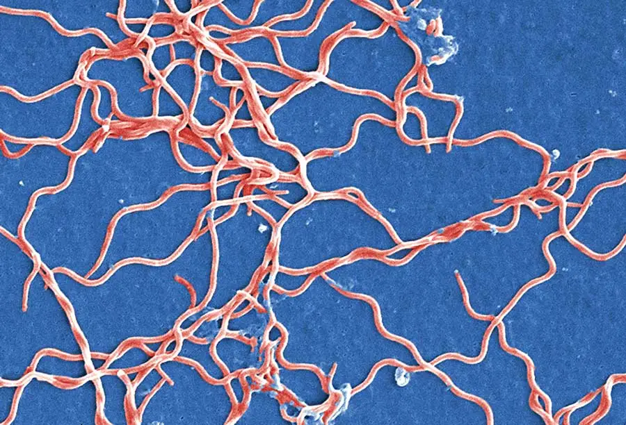
Tick infestations are a recurring roadblock of human development around the world, with estimated damages in the global economic landscape being as high as 30 billion USD. Specifically, India has long been susceptible to tick-borne diseases, due to multiple species invading the livestock. These regional parasites are major vectors for Crimean–Congo hemorrhagic fever virus (CCHFV), a disease with a devastating case fatality rate of 10–40%. While the main way of combatting the infestation of ticks and their carried disease has always been pesticides, often to an invasive degree of their own, scientists are working diligently for ways to produce a vaccine for this deadly and prevalent outbreak. One such method that has been explored is multi-epitopic peptide vaccines that combat Crimean–Congo hemorrhagic fever virus, specifically through the potential immune stimulatory responses they cause.
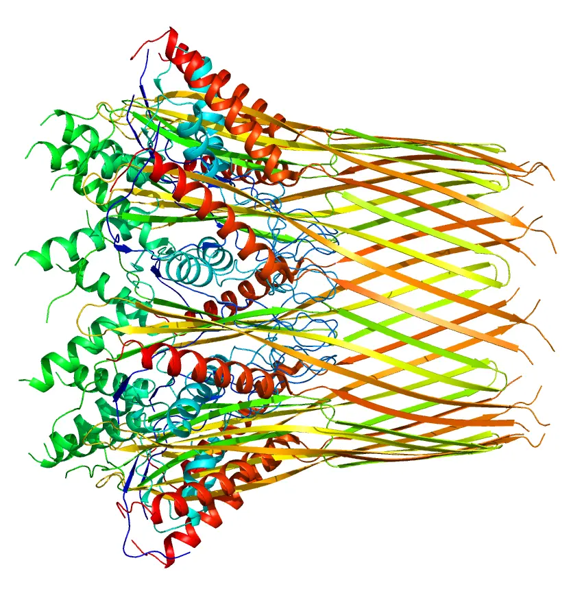
Multi-epitopic peptides help boost immune system
LifeTein provided the group with the two designed multi-epitopic peptides, VT1 and VT2. Using the two peptides, the group put them into two working vaccines in an effort to explore how effective they were at fighting back the ticks. With rabbits, they found strong immunity conferred by the vaccine, displayed by quick larval detachment, delayed tick feeding, low engorgement weights, and overall efficacy against both tick larvae and adults. The results show just how effective treatment with the vaccines are against ticks carrying CCHFV, and the compatibility with rabbits is a great starting point.
In an ideal experiment, the group would have tested on cattle, since that is a much more affected group by these ticks. Regardless, the suitability and stability displayed warrants more attention be put into these multi-epitopic peptide vaccines. Their efficacy displayed against infestations as such is sure to save the global economy billions, as well as countless lives. Immunization in this route is surely more appealing than that of constant and overwhelming pesticides being put in place at every conceivable turn. LifeTein is excited to see where else peptide-based vaccines can be implemented and what other unique properties they can bring to the table.
Nandi A, Manisha, Solanki V, Tiwari V, Sajjanar B, Sankar M, Saini M, Shrivastava S, Bhure SK, Ghosh S. Protective Efficacy of Multiple Epitope-Based Vaccine against Hyalomma anatolicum, Vector of Theileria annulata and Crimean–Congo Hemorrhagic Fever Virus. Vaccines. 2023; 11(4):881. https://doi.org/10.3390/vaccines11040881