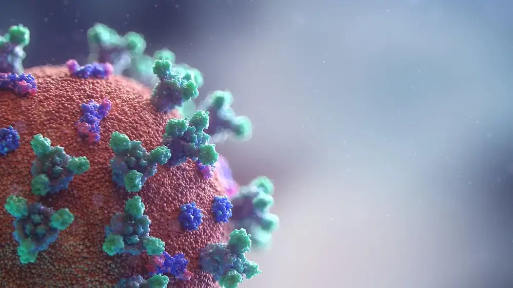
The pivotal role of TMPRSS2 in COVID19

Transmembrane serine protease 2 (TMPRSS2) is a serine protease that in humans is encoded by the TMPRSS2 gene. It is a cell surface protein primarily expressed by endothelial cells across the respiratory and digestive tracts. The protein contains a type II transmembrane domain, a receptor class A domain, a scavenger receptor cysteine-rich domain and a protease domain.

Recent evidence suggested that SARS-CoV-2 uses the ACE2 receptor for cell entry, in synergy with the host’s TMPRSS2. The viral S glycoprotein is cleaved by TMPRSS2, thus facilitating viral activation. As TMPRSS2 is a serine protease, it primes the spike-domain (S) of SARS-CoV-2 by cleaving as the S1/S2 sites. TMPRSS2 activity is crucial for cell entry and viral pathogenesis. In a recent in vitro study by Hoffmann et al., the TMPRSS2 inhibitor camostat mesylate blocked the SARS-CoV-2 entry into primary lung cells, suggesting that TMPRSS2 could represent a potential target in SARS-CoV-2 treatment. This drug is approved for clinical use already in Japan for unrelated illnesses and could serve to be an important therapy for COVID-19.

TMPRSS2 in COVID19
SARS-CoV-2 uses the ACE2 receptor for cell entry, in synergy with the host’s TMPRSS2

Receptor-binding Domains of SARS-CoV-2, LT5578, Cited by Nature

A highly conserved cryptic epitope in the receptor-binding domains of SARS-CoV-2, LT5578, was cited by Nature (Potently neutralizing and protective human antibodies against SARS-CoV-2, volume 584, pages443–449(2020)). This peptide was synthesized in 6 days. This is part of the receptor-binding domain (RBD). It is a critical determinant of virus-receptor interaction and thus of viral host range and tropism. The RBD also includes important viral-neutralizing epitopes (21–23), and it may be sufficient to raise a protective antibody response in inoculated animals.

Two potently neutralizing monoclonal antibodies, COV2-2196 and COV2-2130, which recognize non-overlapping sites, bound simultaneously to the S protein and neutralized wild-type SARS-CoV-2 virus in a synergistic manner.

Lately, a recombinant SARS-CoV-2 Spike S (S1+S2) Protein was produced by LifeTein. The amino acid sequences of recombinant protein was derived from (Q14 – Q1208) of accession# YP009724390.1. The SARS-CoV-2 spike (S) protein is composed of two subunits; the S1 subunit contains a receptor-binding domain that engages with the host cell receptor angiotensin-converting enzyme 2 and the S2 subunit mediates fusion between the viral and host cell membranes. The S RBD protein plays key parts in the induction of neutralizing-antibody and T-cell responses, as well as protective immunity, during infection with SARS-CoV-2 (2019-nCoV) as in recent COVID-19 outbreak.

SARS-CoV-2 receptor binding domain structure
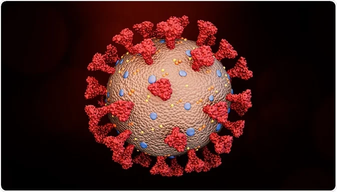
Schematic of the SARS-CoV-2 structure; the illustration of the virus is available at doi: https://doi.org/10.1371/journal.ppat.1008762.g003.

Aureocin A53 is an antimicrobial peptide produced by Staphylococcus aureus A53

LifeTein successfully synthesized Aureocin A53, a highly cationic 51-residue peptide containing ten lysine and five tryptophan residues,   Formyl-MSWLNFLKYIAKYGKKAVSAAWKYKGKVLEWLNVGPTLEWVWQKLKKIAGL, using solid-phase peptide synthesis approach. A53 is a Class II bacteriocins. It was originally isolated from Staphylococcus aureus A53 and is active against methicillin-resistant Staphylococcus aureus (MRSA) and vancomycin-resistant Enterococcus. The mode of action was proposed as insertion into lipid bilayer and consequent membrane leakage. The solid-phase peptide synthesis provide an inexpensive alternative to isolation from bacteria culture or recombinant expression.

Alexa647
Alexa647

Aviptadil Fast-Tracked for Respiratory Distress in COVID-19

It was found that RLF-100 (Aviptadil) is associated with rapid respiratory failure recovery among COVID-19 Patients. The clinical findings may be based on evidence that VIP inhibits the replication of the SARS-CoV-2 virus in human lung cells and immune cells (monocytes). No other antiviral agent has demonstrated rapid recovery from viral infection and demonstrated laboratory inhibition of viral replication. It is a patented formulation of aviptadil (synthetic human Vasoactive Intestinal Polypeptide, VIP), which has been granted FDA fast track designation, FDA emergency use IND authorization, and an expanded access protocol. Aviptadil is an injectable formulation of the vasoactive intestinal polypeptide (VIP) in combination with the adrenergic drug phentolamine.

Aviptadil (Senatek), Vasoactive intestinal polypeptide

Peptide Library: SARS-CoV-2 Receptor Binding Domains

Coronavirus
Coronavirus receptor binding domain. The key receptor binding domain (residues 319-541) is highlighted in yellow. Variable amino acid residues between SARS-CoV-2 and SARS-CoV are highlighted in cyan. Tyr 489, Asn 487, Gln 493, Tyr 505 are important for ACE2 binding.

LifeTein can help in your research with custom peptide synthesis of the following specific proteins: SARS-CoV-2 Receptor Binding Domains, SARS-CoV-2 Nucleocapsid Fragments, T-cell and B-cell Epitopes of SARS-CoV-2, Fusion Inhibitors Targeting HR1 Domain of the SARS-CoV-2 Spike Proteins, Inhibitors of SARS-CoV-2 Mpro/3CLpro/C30 Endopeptidase, ACE2 Inhibitors and Substrates, and AT2 Receptor Agonists and Antagonists.

Pool of 22 peptides derived from a peptide design (15mers with 5 aa overlap) through the receptor binding domain of S1 protein.

Modifications: N-Terminal: Biotin Labeling

Amount: 1mg per peptide

Purity: 95%

Delivery Format: Freeze dried powder

Application(s): Antibody screening, T-cell assays, Immune monitoring, Antigen specific T-cell stimulation, Cellular immune response

Indication(s)/Topic(s): Covid-19, Infection, Respiratory infection

Delivery Time: 2 weeks

SARS-CoV-2 Receptor Binding Domains Overlapping Peptide Pools:

  • QPTESIVRFPNITNL
  • NITNLCPFGEVFNAT
  • VFNATRFASVYAWNR
  • YAWNRKRISNCVADY
  • CVADYSVLYNSASFS
  • SASFSTFKCYGVSPT
  • GVSPTKLNDLCFTNV
  • CFTNVYADSFVIRGD
  • VIRGDEVRQIAPGQT
  • APGQTGKIADYNYKL
  • YNYKLPDDFTGCVIA
  • GCVIAWNSNNLDSKV
  • LDSKVGGNYNYLYRL
  • YLYRLFRKSNLKPFE
  • LKPFERDISTEIYQA
  • EIYQAGSTPCNGVEG
  • NGVEGFNCYFPLQSY
  • PLQSYGFQPTNGVGY
  • NGVGYQPYRVVVLSF
  • VVLSFELLHAPATVC
  • PATVCGPKKSTNLVK
  • TNLVKNKCVNFNFNG

The Cationic Host Defense Peptides Could Be Used To Kill Enveloped Novel Coronavirus SARS-CoV-2

Direct antimicrobial mechanisms of cationic host defense peptides

The cationic host defense peptides (CHDP), also known as antimicrobial peptides, could be used to kill enveloped viruses such as the 2019 Novel Coronavirus SARS-CoV-2. The peptides have the potential to destabilize the viral envelope on contact, damaging the virions and inhibiting infectivity. The specific antiviral peptide may bind to cellular receptors involved in viral infection or peptide-mediated aggregation of viral particles. The antiviral peptides could create an ‘antiviral shield’ at mucosal surfaces and prevent replication and spread of the Coronavirus if upregulated after the initial infection.

During pandemics, where there is insufficient time to produce vaccines (such as the outbreak of respiratory illness Covid-19 first detected in Wuhan, China), the cationic host defense peptides could be the first-line antiviral treatments.

Some of the antimicrobial peptides are the human cathelicidin LL-37 and β-defensins. Cathelicidins are immunomodulatory antimicrobials with an important role in the regulation of the inflammatory response. The only human cathelicidin, LL-37, is the most well-studied peptide in this family. LL-37 is an α-helical peptide. While defensins have a common β-sheet core stabilized with three disulfide bridges between six conserved cysteine residues.

Direct antimicrobial mechanisms of cationic host defense peptides can be mediated by membrane translocation of the peptides followed by binding to intracellular targets such as nucleic acids and/or proteins to kill bacteria. Proline-rich antimicrobial peptides use inner membrane transporters as Trojan horses to gain entry and bind to intracellular targets such as nucleic acids or nascent proteins. And subsequently affect cell processes such as replication, transcription, translation, protein folding, and cell wall synthesis.

At this stage, only a few peptide-derived treatments have made it to market such as PAC-113, a histatin analog, and dalbavancin, a semisynthetic lipoglycopeptide.

Despite the limited understanding of structure-function relationships, the potential of peptide-based therapies remains a promising new clinical direction for the Coronavirus.

2019-nCoV Coronavirus Receptor -Binding Motif Directly Contacts ACE2 Receptor

2019-nCoV Coronavirus Receptor -Binding Motif Directly Contacts ACE2 Receptor
2019-nCoV Coronavirus Receptor -Binding Motif

The extensive structural analyses have revealed that interactions between SARS-CoV spike protein receptor-binding domain (RBD) and its host receptor angiotensin-converting enzyme 2 (ACE2), which regulate both the cross-species and human-to-human transmissions of SARS-CoV.

Studies showed that the sequence of 2019-nCoV coronavirus RBD, including its receptor -binding motif (RBM) that directly contacts ACE2 and uses ACE2 as its receptor with much higher affinity (10-20 times higher!) than SARS.

Several critical residues in 2019-nCoV RBM may provide favorable interactions with human ACE2 such as Gln493 and Asn501.

A total of nine cysteine residues are found in the RBD, six of which forming three pairs of disulfide bonds. Among these three pairs, two are in the core (Cys336-Cys361 and Cys379-Cys432) to help stabilize the β sheet structure while the remaining one (Cys480-Cys488) connects loops in the distal end of the RBM.

LifeTein synthesized a 69 amino acid spike glycoprotein in 6 days

Coronavirus SARS-CoV-2

2019 Novel Coronavirus SARS-CoV-2 is a virus identified as the cause of an outbreak of respiratory illness Covid-19 first detected in Wuhan, China.

To help expedite Covid-19 research, LifeTein synthesized a 69 amino acid spike glycoprotein with one disulfide bond in 6 days. This effort is a partnership with a biotech company for drug development.

Cyclic peptides as broad-spectrum antiviral agents

Cyclic peptides as broad-spectrum antiviral agent

Cyclic peptides as broad-spectrum antiviral agents

Antiviral drugs and vaccines are the most powerful tools to combat viral diseases. Most drugs and vaccines only target a single virus. However, the broad-spectrum antivirals can be used for rapid management of new or drug-resistant viral strains. Cyclized peptides and peptide analogs are excellent examples of broad-spectrum antivirals.

An artificial peptide molecule was found to neutralize a broad range of group 1 influenza A viruses, including H5N1. The peptide design was based on complementarity determining region (CDR) loops have been reported for other viral targets. The optimized peptides bind to the highly conserved stem epitope and block the low pH-induced conformational rearrangements associated with membrane fusion.

These peptidic compounds and their advantageous biological properties should accelerate development of novel small molecule and peptide-based therapeutics against influenza virus.

The linear peptide is Suc-SQLRSLEYFEWLSQ-NH2. Three cyclization strategies were used: head to tail, side chain to side chain and side chain to tail. An ornithine (Orn) side chain was fused with the carboxyl terminus of β-alanine for lactam formation.

Check here for more details: Potent peptidic fusion inhibitors of influenza virus, Science 28 Sep 2017, DOI: 10.1126/science.aan0516

Lately, more broad-spectrum antiviral agents were found to target viruses. It was found that 55 compounds can target eight different RNA and DNA viruses. Dalbavancin is a novel lipo-glycopeptide antibiotic. The lipoglycopeptide disrupts bacterial cell wall formation by binding to
the terminal d-alanyl-d-alanine peptidoglycan sequence in Gram-positive bacteria in a linear, concentration-dependent manner. The dalbavancin has effects on echovirus 1, ezetimibe against HIV1 and Zika virus.

More details: https://www.ncbi.nlm.nih.gov/pubmed/29698664

Magnetic Beads: Expert Tips and Protocols for Effective Use

Magnetic beads

Protein Purification Using Magnetic Beads: Top Tips for Success

Magnetic bead-based protein purification offers a powerful solution for various applications like high-throughput microscale purification, pull-down/CoIP experiments, and protein-protein or protein-DNA interaction studies. Here’s why magnetic beads are the top choice: they can be coated with specific affinity ligands for antigens, antibodies, proteins, or nucleic acids. Moreover, magnetic beads are non-porous and have a defined diameter, eliminating hidden surfaces where molecules can stick, leading to reduced background, simplified purification, and streamlined washing steps. Compared to traditional bead separation methods involving agarose, sepharose, or silica beads, magnetic bead separation stands out as the quickest, cleanest, and most efficient technique.

If you’re new to working with magnetic beads, here are some essential tips to ensure success:

  1. Thorough Resuspension: Ensure uniformity across aliquots by thoroughly resuspending your magnetic beads. These nano-superparamagnetic beads are covalently coated with highly functional groups, providing increased binding capacity and better dispersion. Since magnetic beads are composed of iron oxide and can settle over time, it’s crucial to vortex and resuspend them thoroughly before use to redisperse the beads.
  2. Enhanced Washing: Minimize non-specific binding by increasing the number of washing steps. Whether you’re using ethanol or the recommended wash buffer, make sure to use an adequate volume of wash solution to cover the bead pellet.
  3. Understanding Functional Groups: Different beads are covalently coated with various functional groups like maleimide, primary amine, NHS, carboxylic acid, purified streptavidin, protein A, reduced glutathione, nickel-charged nitrilotriacetic acid, or groups for DNA/RNA purification. These coatings, along with buffer conditions, affect bead properties. Understanding these specifics is essential for proper bead handling.
  4. Efficient Bead Capture: Magnetic beads typically form a pellet attracted to the magnet within a minute. Extend the attraction time to ensure efficient bead capture.
  5. Gentle Supernatant Removal: When removing the wash solution or supernatant, angle the pipette tip to avoid disturbing the magnetic bead pellet. Ensure that the tip doesn’t come into contact with the pellet.

By following these tips, you can make the most of magnetic bead-based protein purification, improving the efficiency and reliability of your experiments.