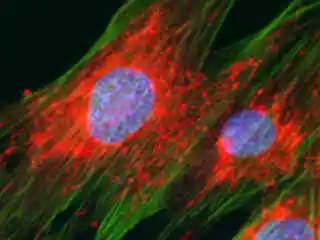
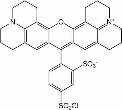
Fluorescent labeling is a powerful technique used in cell biology and microscopy to visualize specific molecules within cells. Among the various fluorophores available, Texas Red stands out as a bright red-fluorescent dye commonly used for cellular imaging applications. It holds many vital applications in biological research.
Texas Red: Properties and Applications
- Bright Fluorescence: Texas Red emits a vibrant red fluorescence when excited by laser lines at 561 or 594 nm. Its brightness makes it ideal for detecting weakly expressed antigens or proteins in biological samples.
- Conjugation to Antibodies and Peptides: Researchers often conjugate Texas Red to antibodies or peptides. When these labeled molecules bind to specific targets (such as antigens), they reveal the location of those targets within cells.
- Photostability: Texas Red exhibits good photostability in buffer and antifade conditions, allowing for reliable imaging over extended periods.
- Alternative: Alexa Fluor 594: For even brighter and more photostable conjugates, consider using Invitrogen Alexa Fluor 594. It shares spectral properties with Texas Red but offers improved solubility and sensitivity.
Find more about Peptide Synthesis here.
Key Takeaways
- Texas Red is a red-fluorescent dye commonly used for cellular imaging.
- It is conjugated to antibodies and proteins to visualize specific targets within cells.
- Researchers can choose between Texas Red and Alexa Fluor 594 based on their specific imaging needs.
Intracellular Localization Studies
Texas Red-labeled antibodies and peptides have been instrumental in studying the localization of specific molecules within cells. By targeting specific antigens or proteins, researchers can visualize their distribution in various cellular compartments:
Nucleus
- Texas Red-conjugated antibodies against nuclear proteins (e.g., histones) allow precise visualization of the nucleus. This aids in understanding chromatin organization and gene expression.
Cytoskeleton
- Texas Red-labeled phalloidin binds to actin filaments, revealing the intricate cytoskeletal network. Researchers use this to study cell motility, shape changes, and intracellular transport.
Membrane Proteins
- Texas Red-labeled antibodies against membrane proteins (e.g., receptors) help identify their presence on the cell surface. This is crucial for signaling studies and drug development.
Multicolor Imaging
Texas Red is often used in multicolor experiments alongside other fluorophores. Combining it with green (e.g., FITC) or blue (e.g., DAPI) fluorophores allows simultaneous visualization of multiple targets within the same sample.
Find more Fluorescents here.
Frequently Asked Questions
- What is Texas Red?
- Texas Red is a red-fluorescent dye commonly used in cell biology and microscopy. It emits bright red fluorescence when excited by specific laser lines, making it ideal for visualizing specific molecules within cells.
- How is Texas Red used in research?
- Intracellular Localization Studies: Texas Red-labeled antibodies and proteins help researchers study the distribution of specific molecules within cellular compartments.
- Can Texas Red be used alongside other dyes?
- Yes, Texas Red can be used in multicolor experiments alongside other fluorophores. Combining it with different colors allows researchers to study multiple targets simultaneously.