Fluorescent Labelling with Cy5

Cy5

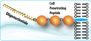
Fluorescent peptide labelling with Cy5, a cyanine dye, has become an indispensable technique in biomedical research, enabling the precise visualization and tracking of peptides in complex biological systems. This method leverages the exceptional photophysical properties of Cy5, which emits in the red to near-infrared region (approximately 650 nm excitation and 670 nm emission), to facilitate a wide range of applications from live-cell imaging to molecular interaction studies. The high molar extinction coefficient and structural versatility of Cyanine5 make it particularly valuable for experiments requiring deep tissue penetration and minimal background autofluorescence, thereby providing enhanced sensitivity and specificity. Consequently, the strategic implementation of Cy5 labelling allows researchers to monitor peptide internalization, investigate protein-protein interactions, and develop advanced diagnostic assays with remarkable clarity and precision.


Key Takeaways

  • Cy5 is characterized by its high molar extinction coefficient and fluorescence in the red to near-infrared spectrum (Ex ~650 nm, Em ~670 nm), which minimizes background interference and is ideal for deep tissue imaging.
  • Common applications include live-cell imagingreceptor internalization studiesFRET-based assays, and the development of sensitive biosensors for pathogen detection.
  • Labelling can be achieved through site-specific methods such as maleimide-thiol coupling or click chemistry, often utilizing a C-terminal cysteine for controlled conjugation.
  • While relatively stable, considerations such as potential photobleaching and the need for efficient purification post-labelling are crucial for maintaining signal integrity and quantitative accuracy.
  • Commercial providers like LifeTein offer comprehensive services for synthesizing labeled peptides, supporting research with a wide array of fluorescent dye options.

Introduction to Cy5 and Its Photophysical Properties

Chemical Characteristics of Cyanine Dyes

Cyanine dyes, including Cy5, belong to a class of synthetic fluorescent molecules characterized by a polymethine bridge connecting two nitrogen-containing aromatic rings. This structure confers a delocalized positive charge, contributing to high extinction coefficients and tunable absorption and emission profiles based on the chain length and chromophores. Cy5, specifically, is a fat-soluble dye that can be modified with sulfonic acid groups to create water-soluble derivatives, enhancing its compatibility with biological systems without significantly altering its optical properties. The dye’s substantial size, however, means it can potentially perturb the biological activity of the labeled peptide, necessitating careful functional validation after conjugation.

Spectral Advantages for Bioimaging

The primary advantage of Cy5 lies in its fluorescence emission in the near-infrared window, which ranges from approximately 650 nm to 670 nm. This spectral range is associated with reduced light scattering and minimal absorption by hemoglobin and water in biological tissues, thereby allowing for deeper penetration and lower background autofluorescence compared to visible light-emitting fluorophores. Consequently, the dye is exceptionally suited for in vivo imaging applications. Furthermore, its compatibility with standard filters for flow cytometry and fluorescence microscopy makes it a versatile choice for various detection platforms.

Find out more about fluorescent peptides here.

Applications of Cy5-Labelled Peptides in Research

Fluorescence Resonance Energy Transfer (FRET)

Cy5 is frequently employed as an acceptor dye in FRET pairs, where it interacts with a donor fluorophore such as Cy3. This configuration is utilized to study protease activityprotein-protein interactions, and conformational changes in peptides. The efficiency of energy transfer in FRET is highly dependent on the proximity between the donor and acceptor, making Cy5-labeled peptides ideal for monitoring molecular interactions and enzymatic cleavage events in real-time. Standardized FRET pairs incorporating the dye are widely available and supported by commercial peptide synthesis services.

Cy5
Cy5-Maleimide

Conjugation Strategies and Practical Considerations

Site-Specific Labelling Techniques

Achieving site-specific conjugation of Cy5 to peptides is essential for preserving biological activity and ensuring reproducible results. Common strategies include:

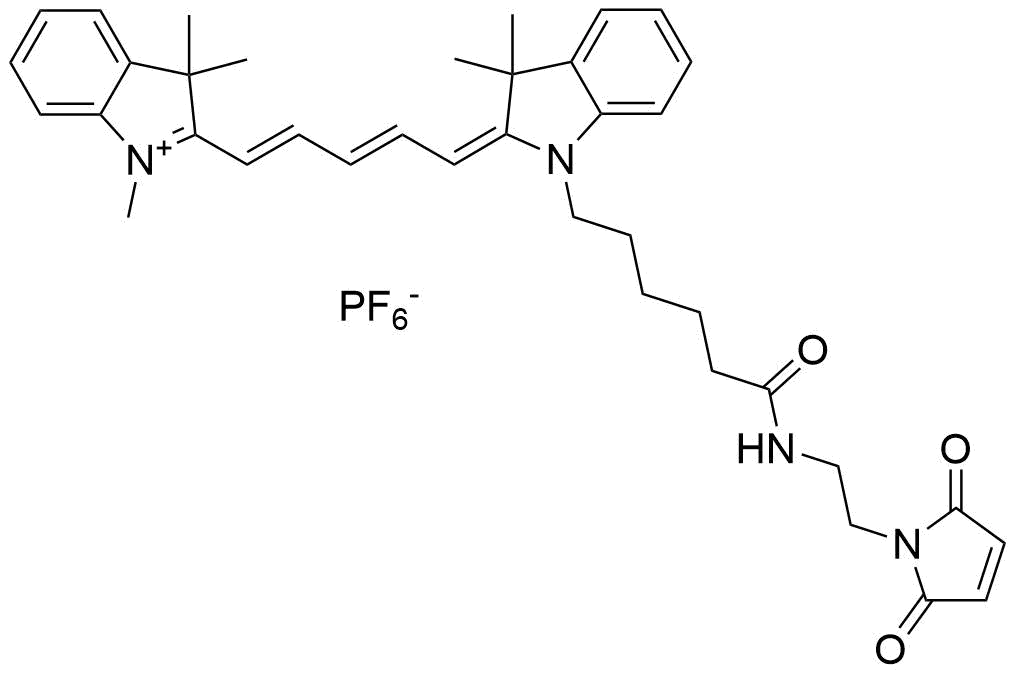
  • Maleimide-thiol chemistry: This method targets cysteine residues, typically introduced at the C-terminus or specific positions within the peptide sequence. The reaction between the maleimide-functionalized Cy5 and the thiol group of cysteine is highly efficient and selective, allowing for controlled labeling with minimal side products.
  • Click chemistry: Copper-catalyzed azide-alkyne cycloaddition (CuAAC) is another robust approach, where an azide-containing peptide reacts with an alkyne-functionalized Cy5 dye. This method offers excellent specificity, compatibility with aqueous buffers, and the ability to label peptides in complex mixtures.
    Additionally, conjugation to primary amines (e.g., on lysine residues or the N-terminus) using NHS ester derivatives of Cy5 is a conventional method, though it may result in heterogeneous labeling if multiple amines are present.

Purification and Validation

Following the conjugation reaction, high-performance liquid chromatography (HPLC) is typically employed to purify the Cy5-labeled peptide from unreacted dye and impurities. Subsequent validation using mass spectrometry (MS) confirms the identity and molecular weight of the conjugate, ensuring labeling efficiency and product correctness. It is also critical to perform functional assays to verify that the Cy5 modification does not adversely affect the peptide’s binding affinity or biological activity, as demonstrated in cAMP accumulation studies for GPCR-targeted peptides.

Find out more about peptide synthesis here.

Frequently Asked Questions (FAQ)

What are the excitation and emission maxima of Cy5?

Cy5 exhibits peak excitation at approximately 650 nm and emission at approximately 670 nm, placing it in the red to near-infrared region of the spectrum. This makes it well-suited for applications requiring minimal background interference and deep tissue penetration.

Can Cy5 be used for in vivo imaging?

Yes, the near-infrared emission properties of the dye make it an excellent fluorophore for in vivo imaging. It allows for non-invasive visualization of biological processes in live animals, such as tumor targeting and biodistribution studies, with high contrast due to reduced absorption by tissue components.

How is Cy5 specifically conjugated to peptides?

Cy5 is typically conjugated using site-specific methods such as maleimide-thiol chemistry (targeting cysteine residues) or click chemistry (via azide-alkyne cycloaddition). These approaches ensure controlled labeling at defined positions, which is crucial for maintaining the peptide’s functional integrity.

Protease OMA1 Activity is Measured by MCA Fluorescent Peptide

MCA Fluorescent Peptide
– Fig. 1. Basis of the OMA1 activity using fluorescence-based peptide.
Fluorescence is released when OMA1 recognizes and cleaves the OPA1 8-mer
peptide (fluorescence reporter) presumably at the RA site, from the cited paper

The continual fission and fusion the Mitochondria undergoes to change its shape and function are a key trait of the organelle, one that is regulated by the enzyme OMA1. However, there is little known regarding OMA1 due to the lack of a consistent method to measure its activity. More information is needed to truly gauge the role of OMA1 as a therapeutic agent. This is where one group sought to measure this activity utilizing a fluorescence-based reporter cleavage assay, one where the protease OMA1 activity is measured by MCA fluorescent peptide.

OMA1 activity measured by (MCA-AFRATDHG-(lys)DNP) peptide

The group arrived at this specific sequence as it includes the specific point on protein OPA1 (between the arginine and alanine) that OMA1 cleaves. They would then be able to spectrofluorometrically measure the fluorescent MCA moiety after the cleavage takes place. The assay proved successful in measuring the activity of OMA1, and in an inexpensive manner. The work clearly lays out the foundation for future studies of OMA1, in both its normal and abnormal pathology.

Julia Tobacyk, Nirmala Parajuli, Stephen Shrum, John P. Crow, Lee Ann MacMillan-Crow, The first direct activity assay for the mitochondrial protease OMA1, Mitochondrion, Volume 46, 2019, Pages 1-5, ISSN 1567-7249, https://doi.org/10.1016/j.mito.2019.03.001.

Revolutionary Antimicrobial Peptides: A New Hope in the Battle Against Citrus Greening

Antimicrobial Peptides

Citrus greening, or Huanglongbing (HLB), is a disease that devastates citrus production all over the world. The culprit behind HLB is the bacterium Candidatus Liberibacter spp. (e.g., CLas), an unculturable pathogen that has proven very difficult to treat. Once a tree is infected, it becomes unproductive and dies within years, costing the global citrus market billions. While current attempts to combat HLB rely on controlling the insect vector, scientists have turned some attention toward the potential of peptides. Their work displayed how antimicrobial peptides show promise for combatting citrus greening, mainly by methods against CLas itself.

Antimicrobial peptides effective against CLas bacteria

With not many current effective options to fight HLB, scientists believe the next area of interest is targeting the CLas secretory pathway using antimicrobial peptides provided by LifeTein. Specifically, the antimicrobial peptides would be blocking the TolC efflux pump protein. The study found three peptides capable of doing this by binding tightly with the TolC receptors and even the β barrel entrance of the protein as well. Treatment with peptides in this manner showed effective inhibition and even mortality in models closely resembling CLas.

The studies displayed using antimicrobial peptides show major promise for future treatment of HLB. With the chemical-resistant bacteria CLas being nearly impossible to slow down, peptides just may have been holding the solution all along. There is hope that new therapies can be developed utilizing the strategies shown, and global citrus production can rest easy after decades of HLB ravaging the farms.

Wang, Haoqi, Nirmitee Mulgaonkar, Samavath Mallawarachchi, Manikandan Ramasamy, Carmen S. Padilla, Sonia Irigoyen, Gitta Coaker, Kranthi K. Mandadi, and Sandun Fernando. 2022. “Evaluation of Candidatus Liberibacter Asiaticus Efflux Pump Inhibition by Antimicrobial Peptides” Molecules 27, no. 24: 8729. https://doi.org/10.3390/molecules27248729

The Vital Role of 14-3-3γ in Influenza A Virus Replication

14-3-3γ

Influenza A is a virus responsible for multiple pandemics over the last centuries, the respiratory disease has claimed millions of lives over the course of human history. Though other pandemics may come to mind in recent years, flu season is just starting up again now as the weather gets chillier. While we can fight back the virus on a yearly basis, more understanding is requited for a long-term victory. A team has been researching into the NS1 protein of the virus, the part responsible for downregulating the antiviral response of host cells to facilitate viral replication. They believe their work has revealed information on the vital role of 14-3-3γ in influenza A virus replication, where the isoform was found to interact with the protein.

Truncated N-Terminus interacts with 14-3-3γ

The group preformed much work such through immunoprecipitation to show the interactions between 14-3-3γ and the influenza A encoded NS1 protein. Some of their most compelling finds was the inhibition of 14-3-3γ expression in the host cells greatly reduced replication of the PR8 wild-type virus, but had no such effect on the R8-NS1/1-98 mutant virus, which lacks most of the effector domain of NS1. LifeTein was able to provide the group with anti β-tubulin antibodies, which assisted in their immunoprecipitation methods.

The team insists that the evidence points directly towards the vital role of 14-3-3γ in influenza A virus replication thanks to the NS1 protein. While they are still unclear on the precise mechanisms of these interactions, they are certain the findings have laid out the groundwork for future pivotal studies involving influenza A and the role of 14-3-3γ in infection.

Kuo, R.-L.; Tam, E.-H.; Woung, C.-H.; Hung, C.-M.; Liu, H.-P.; Liu, H.M.; Wu, C.-C. Interactome Profiling of N-Terminus-Truncated NS1 Protein of Influenza A Virus Reveals Role of 14-3-3γ in Virus Replication. Pathogens 2022, 11, 733. https://doi.org/10.3390/pathogens11070733

Peptides Fold and Self-Assemble on Graphite-Water Interfaces

J. Chem. Inf. Model. 2022, 62, 17, 4066-4082

The concept of self-assembling peptides is a promising front where construction of devices can be achieved through a single molecule. While the outcome is enticing, the means to reach a consistent outcome are complex to say the least. Dozens of factors go into how a peptide may self-assemble and fold, with the most important being the sequence itself. While this can be handled by careful screening and simulations, the interface at which this folding occurs becomes more important to consider at well. Researchers looked to test how specific peptides fold and self-assemble on graphite-water interfaces, where a number of factors give this method the advantage over doing so in free solution.

Graphite helps peptides self fold into conformations

The group studying this phenomenon claimed that the folded conformations of the peptides were stable over a variety of temperatures when observed over graphite. They point out that it is due to the peptide backbone aligning with the zigzag directions of the graphite plane, thus allowing the conformations to occur more favorably from the intermolecular hydrogen bonds of the molecule. Atomic force microscopy revealed these theories to be true beyond initial simulations as well.

The team believes the design principles displayed in these experiments could be of great use in future iterations of self-assembling peptide engineering. The thermodynamically favored self-assembly with the use of a graphite-water interface shows promise as a medium for even more complex molecular devices in the future, a future LifeTein is looking forward to being a part of.

Justin Legleiter, Ravindra Thakkar, Astrid Velásquez-Silva, Ingrid Miranda-Carvajal, Susan Whitaker, John Tomich, and Jeffrey Comer
Journal of Chemical Information and Modeling 2022 62 (17), 4066-4082
DOI: 10.1021/acs.jcim.2c00419

Revolutionary LIPSTIC Method with LPETG Peptide Illuminates Receptor-Ligand Interactions In Vivo And In Vitro

LPETG Peptide
-LIPTSTIC mechanism, from the cited paper.

Cell interaction analysis is a cornerstone of biological research, providing critical insights into the intricate world of molecular communication within living organisms. While traditional microscopy offers a glimpse into these interactions, it often falls short when it comes to revealing the specific receptors and ligands involved. Enter a groundbreaking method known as Labeling Immune Partnerships by SorTagging Intercellular Contacts, or LIPSTIC for short, which has been developed by a team of innovative scientists using LPETG Peptide.

At the heart of LIPSTIC lies the ingenious combination of a fluorescent LPXTG peptide motif and Staphylococcus aureus transpeptidase Sortase A (SrtA), offering a highly effective means of tracking and studying cell interactions. This novel approach is readily detectable through flow cytometry, making it a game-changer in the field of biological research.

The LIPSTIC method hinges on the LPETG peptide and SrtA reaction, a technique that allows for the labeling of receptor and ligand interactions. LifeTein, a leading supplier in the life sciences industry, played a pivotal role by providing the necessary Biotin-ahx-LPETG peptide to the research group. In the LIPSTIC method, a noteworthy ligand or receptor is fused with a tag composed of five N-terminal glycine residues (G5). The SrtA enzyme then graciously donates the fluorescent peptide to this fusion, enabling precise monitoring of the acceptor cell post-separation.

One of the most impressive aspects of LIPSTIC is its versatility. It empowers scientists to analyze cell-cell interactions both in vitro and in vivo, offering a comprehensive understanding of molecular partnerships in various biological contexts. Moreover, LIPSTIC’s sensitivity is a standout feature, as it can even detect rare or low-intensity interactions that might have otherwise remained hidden.

In conclusion, the introduction of the LIPSTIC method marks a significant advancement in the field of cell interaction analysis. Its ability to unveil the intricacies of receptor-ligand interactions in living systems, along with its applicability in diverse research settings, positions LIPSTIC as a powerful tool for scientists striving to unlock the secrets of cellular communication.


Pasqual G, Chudnovskiy A, Tas JMJ, Agudelo M, Schweitzer LD, Cui A, Hacohen N, Victora GD. Monitoring T cell-dendritic cell interactions in vivo by intercellular enzymatic labeling. Nature. 2018 Jan 25;553(7689):496-500. doi: 10.1038/nature25442. Epub 2018 Jan 17. PMID: 29342141; PMCID: PMC5853129.

αCT1 Peptide Weakens Cancerous Glioma Cells

αCT1 Peptide

Glioblastoma (GBM) is the most commonly occurring terminal brain cancer. Due to complications in the brain like the blood brain barrier, methods of treating GBM are few and far between. Therefore, treatment in the region is generally left to specific chemotherapeutics like temozolomide (TMZ), which has the unique capability to bypass the brain blood barrier. However, matters become more complicated as many subpopulations of GBM, namely the glioma stem cell populations, are resistant to TMZ. Researchers are looking into ways to bypass this resilience, namely connexin 43 (Cx43) hemichannels that when inhibited by mimetic peptides allow the glioma stem cell populations to be treated significantly more effectively by TMZ

Cx43 mimetic peptides weaken cancer’s resistance to TMZ

Researchers used LifeTein’s peptide synthesis service to create two mimetic peptides of Cx43, αCT11 and αCT1, to inhibit Cx43 hemichannels and then sensitize the glioma cells and other GBM cell populations to TMZ in a 3D hyaluronic acid and collagen hydrogel-based tumor organoid system. After testing this model extensively, the group found that only the αCT1 peptide in combination with TMZ proved effective in treating the cell lines. It is believed that the αCT1 is more successful due to its cell penetrating sequence when compared to αCT11.

Overall, the group emphasizes that the model used does not accurately mimic the cellular heterogeneity of GBM, but the results are a fantastic start and can be used as a tool to further study treatment of this aggressive brain cancer. Further work can optimize this treatment and can hopefully provide a chance for those who have to go against this fatal ailment.

Jingru Che, Thomas J. DePalma, Hemamylammal Sivakumar, et al. αCT1 Peptide Sensitizes Glioma Cells to Temozolomide in a Glioblastoma Organoid Platform. Authorea. April 29, 2022.

Exploring the Role of Methylated Peptides in Histone Methylation: A LifeTein Perspective

cell-penetration-peptide

Histone methylation, a process that can signal either transcriptional repression or activation, is increasingly recognized for its interrelation with DNA methylation in mammals. For instance, the targeting of DNA methylation is intricately linked to H3K9 methylation, a key regulatory mechanism in gene expression. The p53 gene, known as the guardian of the genome and frequently mutated in human cancers, is regulated by various PTMs, including methylation.

Post-translational modifications (PTMs) of histone proteins, such as acetylation, methylation, and phosphorylation, are pivotal in regulating chromatin dynamics. Among these, the role of methylation, particularly at arginine or lysine residues, stands out for its complexity and significance. LifeTein, a leader in peptide synthesis, has contributed significantly to this field by synthesizing mono-, di-, or tri-methylated peptides. These peptides are instrumental in studying protein-protein interactions, especially in the context of histone methylation.

LifeTein’s contribution to this research is highlighted in a study focusing on the ASHH2 CW domain, which recognizes the methylation state at lysine 4 of histone 3 N-terminal tails. This domain is crucial in recruiting the ASHH2 methyltransferase enzyme to histones. The study utilized H3 histone tail mimicking peptides, specifically monomethylated (ARTK(me1)QTAR), dimethylated (ARTK(me2)QTAR), and trimethylated (ARTK(me3)QTAR) peptides, all synthesized by LifeTein with a remarkable 95% purity as confirmed by mass spectrometry.

The research documented the assignment of a shortened ASHH2 CW construct, CW42, which showed similar binding affinity and better expression yields than previous constructs. This advancement is significant in understanding how different methylation states affect protein-peptide interactions. The study also performed 1H–15N HSQC-monitored titrations to determine the saturation point of the protein-peptide complex. The findings revealed that the CW42 domain, when bound to the monomethylated histone tail mimic, showed similar perturbations in shifts as the di- and tri-methylated instances.

In summary, LifeTein’s synthetic methylated peptides have been instrumental in advancing our understanding of histone methylation. Their high-purity peptides have enabled researchers to delve deeper into chromatin dynamics and gene regulation complexities, paving the way for future discoveries in epigenetic therapies and cancer treatment.

Read the full article on SpringerLink](https://link.springer.com/article/10.1007/s12104-018-9811-x) for more detailed insights into this groundbreaking research.

LifeTein Launches Rush Custom Peptide Synthesis Service: Peptide Delivered in 3-5 Days

Peptide-Library
Peptide-Library

LifeTein is unveiling an expedited peptide synthesis program, promising to place peptides in its customers’ hands within 3-5 business days. The RushPep™ peptide synthesis service was designed to circumvent the existing limitations of conventional solid-phase peptide synthesis (SPPS), which involves a long coupling time and low yield. RushPep™ shortens the time needed for individual coupling, deprotection, and washing steps. The proprietary methodology renders processing ten times faster than in classical synthesis while circumventing the limitations caused by forming by-products or intermediates to which traditional SPPS approaches are subject.

LifeTein’s Rush Custom Peptide Synthesis Service

“When designing the RushPep™ methodology, our focus was not only to produce peptides of high quality and purity but also to offer a streamlined solution that would increase the efficiency of researchers’ protein discovery workflows,” stated Dr. Ya Chen, Head of LifeTein’s Rush Peptide Synthesis Group. “RushPep™ achieves these goals by synthesizing the peptides in 3–5 business days to accelerate research and discovery.”

Chen continued, “The reliability of RushPep™ rush peptide synthesis ensures that the peptides are finished in 3–5 business days with high-batch-to-batch reproducibility. ” Most of the crude peptides have a purity of over 80%. RushPep™ peptide service is valuable for scientists and researchers because it allows them to finish their proteomics projects quickly and cost-effectively.

ID2 peptide for inhibition of tumour growth

Biotinylated wild-type and modified (pT27 and T27W) ID2 peptides (amino acids 14–34) were synthesized by LifeTein. ID2 binds to the VHL ubiquitin ligase complex. This ID2 peptide could be used to inhibit tumor growth in patients with glioblastoma.

LifeTein’s ID2 Peptides Can Inhibit Tumour Growth

Nature, 529, 172–177 (14 January 2016) doi:10.1038/nature16475, An ID2-dependent mechanism for VHL inactivation in cancer.