Featured Product New!: Monoclonal Antibodies to Coronavirus SARS-CoV-2 RBD Domain
Description:Recombinant SARS-CoV-2 Spike S1 Protein
Product: Recombinant protein of severe acute respiratory syndrome coronavirus 2 (SARS-CoV-2) Spike S1, with a rabbit Fc tag at its C-terminus.
Cat #: LTP-V006
Host: Human 293 cells
RefSeq: NC_045512.2; YP_009724390.1; Gene ID: 43740568
Molecular Weight: The amino acid sequences of recombinant protein was derived from the Q14-A684 region of accession# YP_009724390.1. The predicted MW of this product is ~100 kDa, however, it runs higher than 130 kDa on the reduced SDS-PAGE due to a post-translational modification when expressing in mammalian cells.
Applications: Antigens, Western, ELISA and other in vitro binding or in vivo functional assays, and protein-protein interaction studies; For research & development use only!
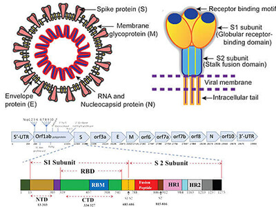
Background: The spike (S) glycoprotein of coronaviruses is known to be essential in the binding of the virus to the host cell at the advent of the infection process. Most notable is a severe acute respiratory syndrome (SARS). The severe acute respiratory syndrome-coronavirus (SARS-CoV) spike (S) glycoprotein alone can mediate the membrane fusion required for virus entry and cell fusion. It is also a major immunogen and a target for entry inhibitors. The SARS-CoV-2 spike (S) protein is composed of two subunits; the S1 subunit contains a receptor-binding domain that engages with the host cell receptor angiotensin-converting enzyme 2 and the S2 subunit mediates fusion between the viral and host cell membranes. The S RBD protein plays key parts in the induction of neutralizing-antibody and T-cell responses, as well as protective immunity, during infection with SARS-CoV-2 (2019-nCoV) as in the recent COVID-19 outbreak.
Quantity: 50ug, Endotoxin level is < 0.1 ng/µg of protein (<1 EU/µg)
Purity: >90% by SDS-PAGE gel and Coomassie Blue staining
Formulation: Purified protein formulated in a sterile solution of PBS buffer, pH7.2, without any preservatives
Download Data File:


Schematic of the SARS-CoV-2 structure; the illustration of the virus is available at doi: https://doi.org/10.1371/journal.ppat.1008762.g003.


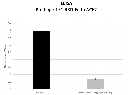
The S1 RBD binds to the ACE2 enzyme, using the Fc only as a negative control, which the RBD did not bind to. The assay was performed with an ELISA, detecting the RBD with streptavidin HRP.