LifeTein Leads the Way in Revolutionary Peptide Conjugation Methods

In the realm of peptide synthesis and bioconjugation, LifeTein stands at the forefront, offering innovative solutions for linking peptides to other biomolecules. Typically, peptides have three biological functional groups available for conjugation: amino (–NH2), carboxyl (–COOH), and thiol (–SH). Among these, the thiol group, particularly from cysteine residues, is often the most effective for bioconjugation. The reaction between maleimides and thiols is a widely recognized method for the bioconjugation and labeling of biomolecules, and LifeTein has mastered this technique to offer superior results.

Click Chemistry: A Revolution in Peptide Conjugation by LifeTein

LifeTein has embraced Click Chemistry, an efficient method for conjugating peptides with various biomolecules. This technique involves modifying the peptide with azide groups (–N3). A standout feature in LifeTein’s arsenal is the novel Copper-free Click Chemistry, which is based on the reaction of a diaryl cyclooctene moiety (DBCO) with an azide-modified peptide. This reaction is not only rapid at room temperature but also avoids the use of cytotoxic Cu(I) catalysts, leading to almost quantitative yields of stable triazoles.

The DBCO component allows copper-free click chemistry to be safely employed with live cells, whole organisms, and non-living samples, which is a significant advantage in various biological applications. Importantly, within physiological temperature and pH ranges, the DBCO group does not react with amines or hydroxyls, which are abundantly present in many biomolecules. The reaction of the DBCO group with the azide group is notably faster than with the sulfhydryl group (–SH, thiol), making it a preferred choice for many of LifeTein’s clients.

Practical Applications: Peptide Drug Conjugations

A prime example of the application of these techniques is in the creation of antibody-biomolecule conjugates. LifeTein’s protocol for Click chemistry of antibody-DNA conjugation is straightforward and efficient:

  1. Pre-conjugation Preparations: Remove all additives from antibody solutions using methods like dialysis or desalting. It’s crucial to eliminate BSA and gelatin from these solutions and concentrate the antibody post-purification.

  2. Activation with DBCO-NHS Ester: The antibody is mixed with a 20-30 fold molar excess of DBCO-NHS ester dissolved in DMSO and incubated at room temperature or on ice.

  3. Quenching the Activation Reaction: This step involves adding Tris-HCl (50-100mM, pH 8) to the reaction mixture, followed by incubation at room temperature or on ice to stabilize the reaction.
  1. Equilibration and Removal of Non-reactive DBCO-NHS Ester: This is achieved using a Zeba column, following the manufacturer’s instructions to ensure precision and effectiveness.

  2. Copper-Free Click Reaction: The DBCO-NHS ester labeled antibody is then mixed with a 2-4 times molar excess of azide-modified oligos. This mixture is incubated overnight at 4°C or for a few hours at room temperature, facilitating the conjugation process.

  3. Validation and Purification: The final step involves validating the conjugation and purifying the product using HPLC, ensuring the high quality and efficacy of the conjugate.

LifeTein’s expertise in peptide synthesis and conjugation is further exemplified by their application of Click Chemistry and thiol-maleimide bioconjugation techniques. These methods are not only efficient but also versatile, opening up new possibilities in the field of peptide-based therapeutics and research.

Selected References:

  • Simon et al. (2012). Facile Double-Functionalization of Designed Ankyrin Repeat Proteins using Click and Thiol Chemistries. Bioconjugate Chem. 23(2):279.
  • Arumugam et al. (2011). [18F]Azadibenzocyclooctyne ([18F]ADIBO): A biocompatible radioactive labeling synthon for peptides using catalyst-free [3+2] cycloaddition. Bioorg. Med. Chem. Lett. 21:6987.
  • Campbell-Verduyn et al. (2011). Strain-Promoted Copper-Free Click Chemistry for 18F Radiolabeling of Bombesin. Angew. Chem. Int. Ed. 50:11117.

Through these advanced techniques, LifeTein continues to be a leader in the field of peptide synthesis and bioconjugation, contributing significantly to the advancement of biomedical research and therapeutic development.

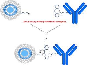
One example of peptide drug conjugations is the antibody-biomoleule conjugate.

click chemistry: DBCO-azide

click chemistry: DBCO-azide

A simple protocol: Click chemistry of antibody-DNA conjugation

Pre-conjugation considerations

  • Remove all additives from antibody solutions using dialysis or desalting.
  • Remove BSA and gelatin from antibody solutions.
  • Concentrate the antibody after dialysis or purification.

Activation of antibodies with DBCO-NHS ester

  • Mix antibody with 20-30 fold molar excess over antibody of DBCO-NHS ester dissolved in DMSO.
  • Incubates at room temperature for 30 min or 2 hours on ice.

Quenching activation reaction

  • Add Tis-Hcl (50-100mM, pH 8) to the reaction.
  • Incubate at RT for 5 min or 15 minutes on ice.

Equilibration and removal of non-reactive DBCO-NHS ester by Zeba column (Follow the manufacturer’s instruction)

Copper-Free click reaction

  • Mix DBCO-NHS ester labeled antibody with 2-4 times molar excess of azide-modified Oligos.
  • Incubated overnight (around 10-12 hours) at 4°C or 3-4 hours at room temperature.

Validation of conjugation and purification by HPLC

A Simple Protocol to Refold Peptides or Small Proteins

Understanding the process of peptide folding is a critical first step toward understanding protein folding. Depending on the temperature and solvent conditions, peptides are highly flexible and can adopt a variety of conformations in solution. Many unfolded peptides could spontaneously refold in vitro to form a native protein with full biological activity in the absence of other factors. Peptide fragments of proteins often have intrinsic propensities for the formation of their native conformations.

Peptide Folding

Proteins are the workhorses inside living cells. The interactions among proteins are critical for various important biological processes. Almost about 15-40% of the protein-protein interactions are peptide-mediated. A short stretch of amino acid residues from one protein partner contributes most to its binding to the other protein structure. These short linear interacting motifs can be found embedded inside disordered regions of intrinsically disordered proteins, or appear as flexible linkers connecting function regions and as flexible loops to rigid fragments and domains.

The primary sequence contains all the information to define the three-dimensional structure of a protein and its biological functions. The mutation or deletion of any amino acid may have a big impact on folding and stability. It takes nanoseconds (ns) for the peptide to form an intermolecular contact. The timescale of loop closing is 10 nanoseconds (ns). The formation of alpha-helical peptides is 200 ns,  beta hairpins, and mini-proteins in 1–10 ms timescale. Many studies had a very good agreement between measured and calculated folding rates. Many factors such as temperature, pH,  molecular chaperones, salts, and denaturants may affect a peptide in reaching its native state.

So it is critical to minimize factors that affect protein refolding. A successful folding should have inadequate denaturant concentrations to destabilize the native state of a peptide or protein. GuHCL can be used in order to disrupt the hydrophobic interactions within the tertiary structure.

  1. The peptide was solubilized in resuspension buffer (50 mM Tris, pH 8, 6 M GuHCl (Sigma, G4505), 10 mM DTT, 2mM EDTA) by vortexing.
  2. Use enough resuspension buffer such that the final peptide concentration is 0.2mg/ml.
  3. The resuspended peptide was then diluted 50% in dialysis buffer #1 (50 mM Tris, pH 8, 2 M GuHCl, 2mM EDTA) resulting in a 4 M GuHCl-containing solution.
  4. The peptide solution was then dialyzed overnight at 4°C in snakeskin dialysis tubing (Pierce) against 2 L of buffer #1.
  5. The following day the dialysis buffer was changed to 2 L of dialysis buffer #2 (50 mM Tris, pH 8, 1 M GuHCl, 0.4 M Arginine (Sigma, A5006), 3 mM Reduced Glutathione, 0.9 mM Oxidized Glutathione, 2mM EDTA) for overnight dialysis at 4°C.
  6. The following day the dialysis buffer was diluted 50% with water and dialysis continued overnight.
  7. Any insoluble material was centrifuged (18000×g at 2–8°C for 20 minutes) and the remaining peptide solution dialyzed overnight at 4°C against 1 L of dialysis buffer #3 (50 mM Tris, pH 8, 250 mM NaCl, 0.1 M Arginine, 3 mM Reduced Glutathione, 0.9 mM Oxidized Glutathione, 2mM EDTA) to remove the remaining GuHCl.
  8. The final dialyzed protein solution was clarified by centrifugation (18000×g at 2–8°C for 20 minutes) and the supernatant was separated by RP-HPLC.

References:

Protein Refolding for Western Blotting

For refolding, proteins in the SDS-Polyacrylamide gels were incubated in transfer buffer I (0.01% Triton X-100, 48 mM Tris, 39 mM Glycine, 20%methanol, pH 9.2) twice for 15 min, and then transfer buffer II (48 mM Tris, 39 mM Glycine, 20%methanol, pH9.2) twice for 15 min, then transferred onto Immobilon-P membranes (Millipore) in transfer buffer II and processed by standard procedures for Western blots.

Reference: Zhou J, Blissard GW. Mapping the conformational epitope of a neutralizing antibody (AcV1) directed against the AcMNPV GP64 protein. Virology. 2006 Sep 1;352(2):427-37. doi: 10.1016/j.virol.2006.04.041. Epub 2006 Jun 14. PMID: 16777166; PMCID: PMC3767133.

Peptide Synthesis Home Page

Our Services:

COVID-19 Services & Products

Custom Antibody Services

Rush Peptide Synthesis

Peptide Nucleic Acids (PNAs)

Custom Peptide Synthesis Services

Gene Synthesis Service

Custom Chemical Synthesis

Other Posts: To Make Simpler and Better Biosensors LifeTein Peptides Used for Pulldown Assay Peptide Applications
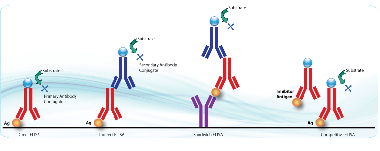
Synthetic Peptides Used for indirect ELISA

ELISA

Peptides can be used for ELISA assay. A peptide-based indirect ELISA was used to screen a population of 40 Multiple sclerosis patients and 39 healthy controls. The encephalitogenic myelin oligodendrocyte glycoprotein (MOG)35–55 synthetic peptides were synthesized by LifeTein.

LifeTein’s Synthetic Peptides for ELISA

All MOG peptides or Mycobacterium avium subspecies paratuberculosis (MAP) peptides were synthesized at >90% purity by LifeTein to make sure the ELISA results are clean and consistent. The plates were coated with peptides. After overnight incubation, the plate was blocked, rinsed, and late react with antibodies according to the protocol. In silico analysis identified two peptides belonging to MAP and BCG, which share sequence homology with MOG(35–55). The peptide-based indirect ELISA data showed that sharing of highly conserved linear amino acidic sequences is necessary to elicit antibody-mediated cross-reactivity.

These findings concluded that the presence of MOG (35–55)-specific antibodies in multiple sclerosis pathogenesis. This can be used as a diagnostic biomarker in multiple sclerosis.

 [PDF] Evaluation of the humoral response against mycobacterial peptides, homologous to MOG35–55, in multiple sclerosis patients

MG Marrosu, LA Sechi – 2014

 All peptides were synthesized at N90% purity commercially (LifeTein, South
Plainfield, NJ 07080 US). Purified peptides were prepared as [10 mM] stock solutions,
and were stored in single- use aliquots at −80 °C. 2.3. ELISA 

Peptide Synthesis Home Page

Our Services:

COVID-19 Services & Products

Custom Antibody Services

Rush Peptide Synthesis

Peptide Nucleic Acids (PNAs)

Custom Peptide Synthesis Services

Gene Synthesis Service

Custom Chemical Synthesis

Other Posts:

LifeTein Peptide Cited in Cell

Phospho-specific antibodies by LifeTein published in Nature

Synthesis of multiple antigenic peptides: strategies and limitations

The Structural Basis of Peptide-Protein Binding Strategies